Review
. 2021 Jul;37(7):1039-1050.
doi: 10.1007/s12264-021-00693-w. Epub 2021 Apr 29.

Oligodendroglial GABAergic Signaling: More Than Inhibition!

Affiliations
Review

Oligodendroglial GABAergic Signaling: More Than Inhibition!

Xianshu Bai et al. Neurosci Bull. 2021 Jul.

Abstract

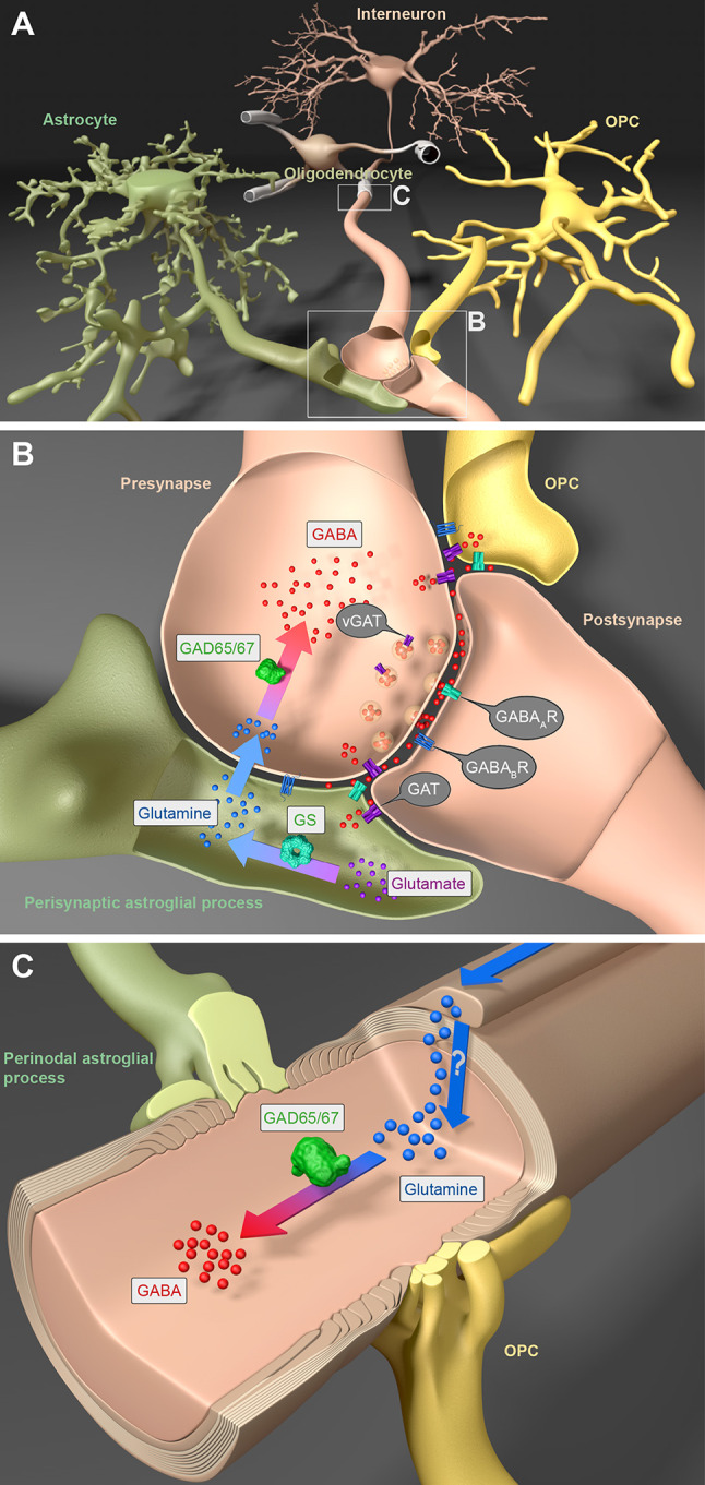
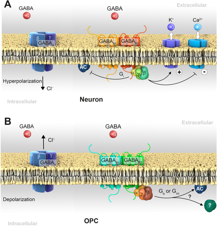
GABA is the main inhibitory neurotransmitter in the CNS acting at two distinct types of receptor: ligand-gated ionotropic GABAA receptors and G protein-coupled metabotropic GABAB receptors, thus mediating fast and slow inhibition of excitability at central synapses. GABAergic signal transmission has been intensively studied in neurons in contrast to oligodendrocytes and their precursors (OPCs), although the latter express both types of GABA receptor. Recent studies focusing on interneuron myelination and interneuron-OPC synapses have shed light on the importance of GABA signaling in the oligodendrocyte lineage. In this review, we start with a short summary on GABA itself and neuronal GABAergic signaling. Then, we elaborate on the physiological role of GABA receptors within the oligodendrocyte lineage and conclude with a description of these receptors as putative targets in treatments of CNS diseases.

Keywords: GABA; GABAA receptor; GABAB receptor; OPC; Oligodendrocyte lineage.


Conflict of interest statement

The authors declare no competing interests.

Figures

Fig. 1
Fig. 1
GABA cycling between interneurons, cells of the oligodendrocyte (OL) lineage, and astrocytes. A In the central nervous system, interneurons form an intricate signaling network with cells of the OL lineage, i.e., myelinating OLs and their precursors (OPCs), and with perisynaptic as well as perinodal processes of astrocytes. B In the synaptic microenvironment, extracellular glutamate is converted into glutamine in astrocytes by glutamine synthetase (GS). After release, glutamine is taken up by interneurons and transformed into GABA by the glutamate decarboxylases GAD65 and/or GAD67. Upon action potential arrival, GABA is released into the synaptic cleft by vesicles expressing GABA transporters (vGAT). After binding to postsynaptic neuronal GABAA and/or GABAB receptors, GABA induces postsynaptic neuronal hyperpolarization. But neuron-released GABA can also act on the GABA receptors of OPCs modulating axonal myelination. In addition, extrasynaptic GABA is taken up by neuronal GAT1 and astroglial GAT3 transporters. Both transporters, however, are also expressed by OPCs, but functional studies are still required to determine their roles. C Also, OLs can express GS to produce glutamine. The latter might be transported to myelinated axons, where it can be converted into GABA. Additional experiments are still required to test this hypothesis.
Fig. 2
Fig. 2
GABA receptor expression in neurons and OPCs. A Activation of ionotropic GABAA receptors induces Cl influx to hyperpolarize neurons. The GABAB1 subunit confers ligand-binding, while the B2 subunit transduces the GABA signal into the cell. Activation of the neuronal GABAB receptor induces dissociation of Gα and Gβγ subunits. The Gα subunit inhibits adenylyl cyclase (AC), while Gβγ activates G protein-gated inwardly rectifying K+ channels and inhibits voltage-gated Ca2+ channels (VGCCs), thereby reducing neurotransmitter release. The regulation of VGCCs can occur pre- and postsynaptically. B Different from neurons, in OPCs, activation of GABAA receptors causes a Cl efflux and depolarization based on the higher levels of cytosolic Cl. GABAB receptors expressed in OPCs are thought to transduce signals via Gα with or without association of Gβγ; or via the Gq pathway linked to phospholipase C, further increasing intracellular Ca2+ release from the endoplasmic reticulum.
Fig. 3
Fig. 3
Synaptic and non-synaptic neuron-OPC communication. A Schematic of neuron-OPC communication in the brain, including direct soma-soma (A1) and synaptic contact (A2). B–D OPC somata (PDGFRα+, red) are in close contact with neuronal somata (NeuN+, green) (arrows) in cortex (ctx, B and C) and hippocampus (hc, D). Micrographs in B and C are from the cortex of NG2-CreERT2 × Rosa26-CAG-lsl-tdTomato mice [6, 133]. Images were acquired by confocal laser-scanning (LSM710, B and C) or automated epifluorescence microscopy (AxioScan.Z1) (D) with appropriate filters and objectives. Scale bars, 20 μm for B and 50 µm for D.

References

    1. Lee SE, Lee Y, Lee GH. The regulation of glutamic acid decarboxylases in GABA neurotransmission in the brain. Arch Pharm Res. 2019;42:1031–1039. - PubMed
    1. Roberts E, Frankel S. gamma-Aminobutyric acid in brain: its formation from glutamic acid. J Biol Chem. 1950;187:55–63. - PubMed
    1. Krnjević K, Schwartz S. The action of gamma-aminobutyric acid on cortical neurones. Exp Brain Res. 1967;3:320–336. - PubMed
    1. Hösli L, Andrès PF, Hösli E. Neuron-glia interactions: indirect effect of GABA on cultured glial cells. Exp Brain Res. 1978;33:425–434. - PubMed
    1. Nishiyama A, Komitova M, Suzuki R, Zhu X. Polydendrocytes (NG2 cells): multifunctional cells with lineage plasticity. Nat Rev Neurosci. 2009;10:9–22. - PubMed

LinkOut - more resources