. 2021 Apr 30;17(4):e1009550.
doi: 10.1371/journal.ppat.1009550. eCollection 2021 Apr.

Salmonella enters a dormant state within human epithelial cells for persistent infection

Affiliations

Salmonella enters a dormant state within human epithelial cells for persistent infection

Chak Hon Luk et al. PLoS Pathog. .

Abstract

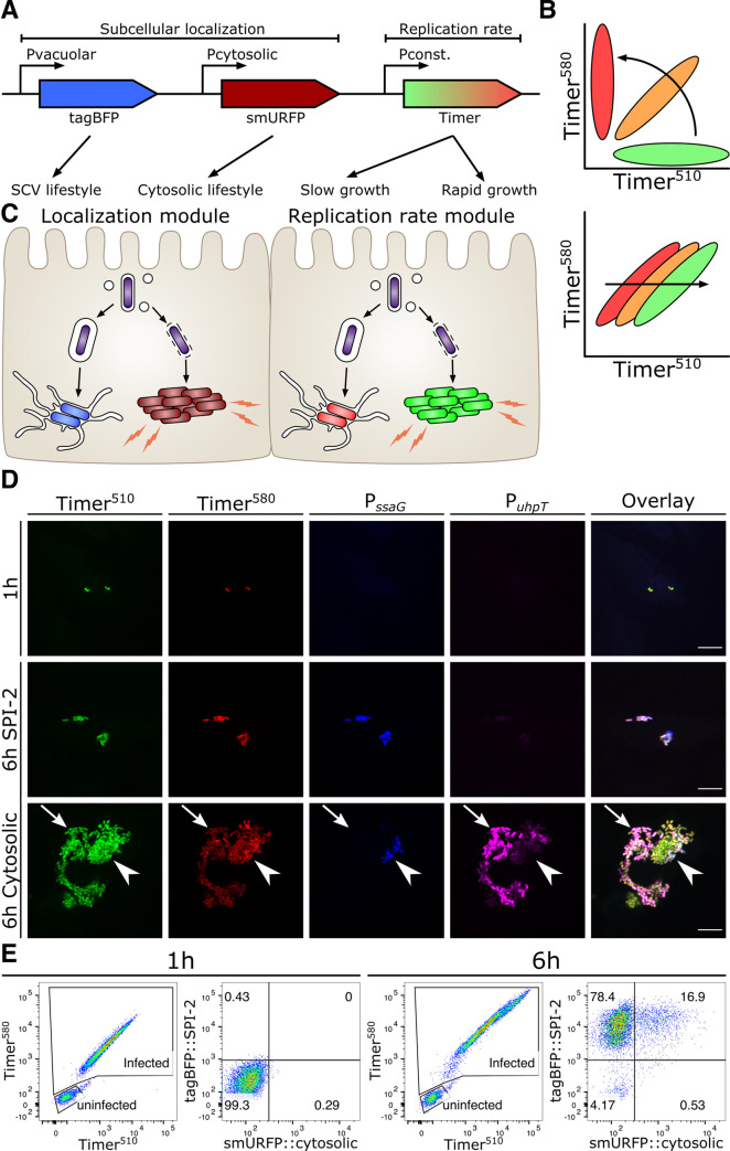
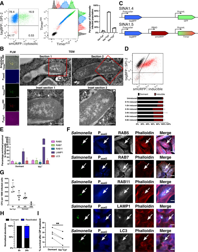
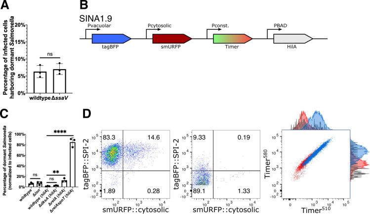
Salmonella Typhimurium (S. Typhimurium) is an enteric bacterium capable of invading a wide range of hosts, including rodents and humans. It targets different host cell types showing different intracellular lifestyles. S. Typhimurium colonizes different intracellular niches and is able to either actively divide at various rates or remain dormant to persist. A comprehensive tool to determine these distinct S. Typhimurium lifestyles remains lacking. Here we developed a novel fluorescent reporter, Salmonella INtracellular Analyzer (SINA), compatible for fluorescence microscopy and flow cytometry in single-bacterium level quantification. This identified a S. Typhimurium subpopulation in infected epithelial cells that exhibits a unique phenotype in comparison to the previously documented vacuolar or cytosolic S. Typhimurium. This subpopulation entered a dormant state in a vesicular compartment distinct from the conventional Salmonella-containing vacuoles (SCV) as well as the previously reported niche of dormant S. Typhimurium in macrophages. The dormant S. Typhimurium inside enterocytes were viable and expressed Salmonella Pathogenicity Island 2 (SPI-2) virulence factors at later time points. We found that the formation of these dormant S. Typhimurium is not triggered by the loss of SPI-2 effector secretion but it is regulated by (p)ppGpp-mediated stringent response through RelA and SpoT. We predict that intraepithelial dormant S. Typhimurium represents an important pathogen niche and provides an alternative strategy for S. Typhimurium pathogenicity and its persistence.


Conflict of interest statement

The authors have declared that no competing interests exist.

Figures

Fig 1
Fig 1. SINA enables precise determination of the different Salmonella intracellular lifestyles in human epithelial cells.
(A) Schematic diagram of the construction of subcellular localization and replication rate modules of SINA1.1. The subcellular localization module is composed of the vacuolar submodule (PssaG-tagBFP) and cytosolic submodule (PuhpT-smURFP), while the replication rate module is composed of a constitutively expressed Timerbac (PybaJ-Timerbac) (B) (Top) Schematic diagram of the emission spectrum shifts of S. Typhimurium harboring Timerbac as Timerbac matures, where emission shifts from green to red (Bottom) Green:Red ratio increases with elevating S. Typhimurium replication rates. As S. Typhimurium divides, both Timer510 and Timer580 fluorophores are diluted. With a higher production rate of Timer510 than Timer580, fast dividing S. Typhimurium exhibits a higher Green:Red ratio. (C) Expected output by SINA as S. Typhimurium dwells in distinct subcellular localizations. Vacuolar S. Typhimurium are of lower replication rate (i.e. lower Green:Red ratio) and are expected to emit blue fluorescence; cytosolic S. Typhimurium are of higher replication rate (i.e. higher Green:Red ratio) and are expected to emit far red fluorescence (D) HeLa cells infected by S. Typhimurium harboring SINA1.1. Output of SINA from intracellular S. Typhimurium was detected by fluorescence microscopy at 1 h pi, vacuolar (arrowhead) and cytosolic (arrow) S. Typhimurium at 6 h pi. (3 independent experiments). Scale bars are 10 μm. (E) HeLa cells infected by S. Typhimurium harboring SINA1.1. Output of SINA from intracellular S. Typhimurium at 1 h and 6 h pi was detected by flow cytometry (3 independent experiments).
Fig 2
Fig 2. S. Typhimurium displays a novel inactive intracellular lifestyle in epithelial cells.
(A) (Left and Middle) Timerbac profile and distribution of single cells with no infection (black), infected cells with inactive bacteria (Vac-Cyt-) (red), infected cells with only vacuolar bacteria (Vac+Cyt-) (blue) and infected cells with both vacuolar and cytosolic populations (Vac+Cyt+) (green) at 6 h pi. (Right) Abundance of S. Typhimurium-infected cells (Vac-Cyt-, Vac+Cyt- and Vac+Cyt+) as illustrated in (A) (n = 3). (B) (Left) Brightfield and fluorescent microcopy (FLM) images of infected HeLa cells harboring Vac-Cyt- S. Typhimurium at 6 h pi. (Right) Serial sections of TEM images of Vac-Cyt- S. Typhimurium, arrowhead indicates host membrane structures of the SCV. (C) Schematic illustration for the constructions of SINA derivatives, SINA1.4 and SINA1.5. SINA1.4 was used for immunofluorescence staining against RAB5, RAB7, RAB11, LAMP1 and LC3; SINA1.5 was used for arabinose induction assay. (D) (Top) Responsiveness of intracellular S. Typhimurium towards an arabinose pulse between 5–6 h pi, uninduced control (black); arabinose-induced (red). (Bottom) Quantification on the responsiveness of Vac- S. Typhimurium pulsed at different time intervals during the infection time course, dormant (black), inducible (maroon). Samples were all harvested at 6 h pi. (n = 3) (E) HeLa cells were infected with SINA1.4-harboring S. Typhimurium, harvested at 6 h pi, fixed and stained. Quantification of the presence of RAB5, RAB7, BAB11, LAMP1 and LC3 proximal to Vac- and Vac+ S. Typhimurium at 6 h pi. (n = 3) (F) Representative images of Vac- S. Typhimurium (arrow) quantified in (D); S. Typhimurium (green), Vac- (blue), RAB5, RAB7, RAB11, LAMP1 and LC3 (grey), Phalloidin (red). (G) Designated populations of infected HeLa cells were enriched by cell sorting and plated for CFU. Quantification of CFU from dormant S. Typhimurium at 6 h, 24 h and 168 h pi and Vac+Cyt- S. Typhimurium at 6 h pi. (n = 5 for Vac+Cyt- 6 h, Dormant 6 h, 24 h; n = 3 for Dormant 168 h) (H) Quantification of SPI-2 activity using flow cytometry in enriched dormant S. Typhimurium at 6 h and enriched Vac-Cyt- infected cells re-plated until 24 h pi. (n = 3) (I) Survival percentage of dormant and Vac+Cyt- intracellular S. Typhimurium against 3 h of CIP treatment, infected cells were harvested at 6 h pi, enriched by cell sorting and plated for CFU. (n = 4) At least a total of 1000 events of infected cells were analyzed by flow cytometry or 50 infected cells by microscopy in each experiment replicates. The bars represent the mean, statistics were performed using unpaired t test (**p<0.01).
Fig 3
Fig 3. S. Typhimurium dormancy is negatively regulated by SpoT.
(A) HeLa cells were infected with SINA1.1-harboring S. Typhimurium, the abundance of Vac-Cyt- population in wild type and SPI-2 mutant ΔssaV infected cells were quantified with flow cytometry at 6 h pi. (n = 3) (B) Schematic diagram for the construction of SINA derivative, SINA1.9, yielded from the introduction of an arabinose-inducible hilA expression cassette into SINA1.1. SINA1.9 was used to rescue the reduced invasiveness of ΔdksA, ΔrelA and ΔrelAspoT mutant strains. (C) HeLa cells were infected with SINA1.1 or SINA1.9-harboring S. Typhimurium, the abundance of Vac-Cyt- population in (p)ppGpp biogenesis and regulon mutants, Δlon, ΔdksA, ΔrelA and ΔrelAspoT were quantified by flow cytometry at 6 h pi (n = 3) (D) Distribution of Vac-Cyt-, Vac+Cyt- and Vac+Cyt+ populations in hilA-expressing wild type (Left) and ΔrelAspoT mutant (Middle) infected HeLa cells at 6 h pi quantified by flow cytometry. Overlay Timerbac profile (Right) of Vac-Cyt- (red) and Vac+Cyt- (blue) populations of wild type and Vac-Cyt- population of ΔrelAspoT mutant (grey) in infected HeLa cells quantified by flow cytometry at 6 h pi. (3 independent experiments) At least a total of 1000 events of infected cells were analyzed by flow cytometry in triplicate experiments. Statistics were performed using unpaired t test. ns: not significant (P > 0.05), **P < 0.01, ****P < 0.0001.
Fig 4
Fig 4. Schematic illustration of the role of (p)ppGpp alarmone pathway on S. Typhimurium dormancy in enterocytes and the proposed pathophysiological implication of S. Typhimurium dormancy in enterocytes.
(A) Schematic diagram of S. Typhimurium lifestyles and the regulatory role of SpoT on S. Typhimurium dormancy in human epithelial cells. S. Typhimurium can opt for three distinct lifestyles: cytosolic, vacuolar and dormant, which exhibits discernible subcellular localization, replication rate and metabolism. The entry of dormant state is negatively regulated by (p)ppGpp synthatase SpoT, while the regulatory mechanism on the dormancy exit remains to be determined. (B) Schematic diagram of S. Typhimurium infection progression in the gut epithelium. As S. Typhimurium reaches the intestinal epithelium, a portion of S. Typhimurium expresses T3SS1 (purple) to enter host cells and adopts various intracellular lifestyles. Distinct S. Typhimurium lifestyles support rapid tissue colonization and gut inflammation to increase competitiveness of luminal S. Typhimurium (red). (Top) Reactivation of dormant S. Typhimurium leads to prolonged gut inflammation that supports the continuous growth of S. Typhimurium at gut lumen. (Bottom) Dormant S. Typhimurium reactivates after the eradication of gut S. Typhimurium, which serves as the reservoirs of infection relapse.

References

    1. Stanaway JD, Parisi A, Sarkar K, Blacker BF, Reiner RC, Hay SI, et al.. The global burden of non-typhoidal salmonella invasive disease: a systematic analysis for the Global Burden of Disease Study 2017. Lancet Infect Dis. 2019;19: 1312–1324. 10.1016/S1473-3099(19)30418-9 - DOI - PMC - PubMed
    1. Shekhar C. International Conference on Food Security and Sustainable Agriculture Global impact of salmonellosis on health and economy. ~ 93 ~ J Pharmacogn Phytochem. 2018;4.
    1. Ilyas B, Tsai CN, Coombes BK. Evolution of Salmonella-host cell interactions through a dynamic bacterial genome. Frontiers in Cellular and Infection Microbiology. Frontiers Media S.A.; 2017. p. 428. 10.3389/fcimb.2017.00428 - DOI - PMC - PubMed
    1. Dandekar T, Astrid F, Jasmin P, Hensel M. Salmonella enterica: a surprisingly well-adapted intracellular lifestyle. Front Microbiol. 2012;3: 164. 10.3389/fmicb.2012.00164 - DOI - PMC - PubMed
    1. Lou L, Zhang P, Piao R, Wang Y. Salmonella Pathogenicity Island 1 (SPI-1) and Its Complex Regulatory Network. Frontiers in Cellular and Infection Microbiology. Frontiers Media S.A.; 2019. p. 270. 10.3389/fcimb.2019.00270 - DOI - PMC - PubMed

Publication types

MeSH terms

Substances

LinkOut - more resources