. 2021 May;31(3):480-492.
doi: 10.1111/jon.12868. Epub 2021 Apr 30.

MRI characteristics of Japanese macaque encephalomyelitis: Comparison to human diseases

Affiliations

MRI characteristics of Japanese macaque encephalomyelitis: Comparison to human diseases

Ian J Tagge et al. J Neuroimaging. 2021 May.

Abstract

Background and purpose: To describe MRI findings in Japanese macaque encephalomyelitis (JME) with emphasis on lesion characteristics, lesion evolution, normal-appearing brain tissue, and similarities to human demyelinating disease.

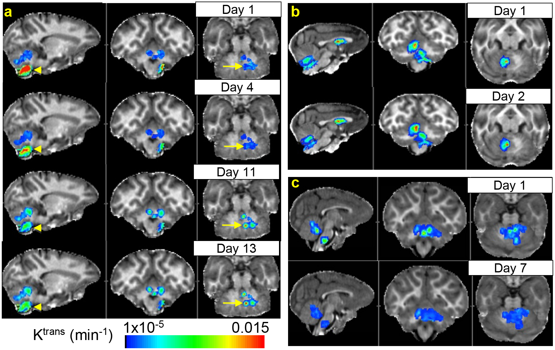
Methods: MRI data were obtained from 114 Japanese macaques, 30 presenting neurological signs of JME. All animals were screened for presence of T2 -weighted white matter signal hyperintensities; animals with behavioral signs of JME were additionally screened for contrast-enhancing lesions. Whole-brain quantitative T1 maps were collected, and histogram analysis was performed with regression across age to evaluate microstructural changes in normal appearing brain tissue in JME and neurologically normal animals. Quantitative estimates of blood-brain-barrier (BBB) permeability to gadolinium-based-contrast agent (GBCA) were obtained in acute, GBCA-enhancing lesions. Longitudinal imaging data were acquired for 15 JME animals.

Results: One hundred and seventy-three focal GBCA-enhancing lesions were identified in 30 animals demonstrating behavioral signs of neurological dysfunction. JME GBCA-enhancing lesions were typically focal and ovoid, demonstrating highest BBB GBCA permeability in the lesion core, similar to acute, focal multiple sclerosis lesions. New GBCA-enhancing lesions arose rapidly from normal-appearing tissue, and BBB permeability remained elevated for weeks. T1 values in normal-appearing tissue were significantly associated with age, but not with sex or disease.

Conclusions: Intense, focal neuroinflammation is a key MRI finding in JME. Several features of JME compare directly to human inflammatory demyelinating diseases. Investigation of JME combined with the development and validation of noninvasive imaging biomarkers offers substantial potential to improve diagnostic specificity and contribute to the understanding of human demyelinating diseases.

Keywords: animal models; blood-brain-barrier; magnetic resonance imaging; neurodegeneration; neuroinflammation.


Figures

Figure 1.
Figure 1.
1H2O T1 values and age in JM Brain. Whole-brain WM and GM T1 average values are plotted as a function of age for 113 JMs (84 HC, 29 JME). No significant T1 differences between JME and HC groups or sex were found. Solid lines represent regression results T1(age) = β0 + β1×age + β2×age2 and dotted lines represent 95% confidence intervals.
Figure 2.
Figure 2.
JME Lesion Locations and Prevalence. Total lesion counts across all animals in each structure are given in panels (a,c). Panels (b,d) illustrates the number of animals presenting with at least one lesion, and its location. Regions are separated by hemisphere where applicable to assess hemispheric preference. R – right; L – left; CBLL – cerebellum; IC – internal capsule; PFWM – prefrontal white matter; PVWM – periventricular white matter; CWM – cerebral white matter; CC – corpus callosum; SC – cervical spinal cord.
Figure 3.
Figure 3.
Incidental WMSH (yellow arrows) on T2-w images in nominally healthy control JM (a: 16 year old female; b: 14 year 7 month old female; c: 16 year old female). The spatial distribution of WMSHs is consistent with that observed in JME lesions and could indicate prior sub-clinical disease activity that resolved without overt or observed behavioral changes.
Figure 4.
Figure 4.
Chronic non-enhancing JME lesions. T2-w and R1 pre- and post-GBCA images in the same axial plane are shown for one case (JM 34678) demonstrating bilateral WMSH corresponding to non-enhancing R1 hypointensities (arrows), consistent with burned-out (non-enhancing) chronic lesions observed in MS.
Figure 5.
Figure 5.
Extensive spinal cord and corpus callosum (CC) lesions in JME observed on proton density weighted images. Longitudinally extensive spinal cord lesions appear bright in three separate cases in panels a, b, and d (JMs 31869 (a), 32150 (b), and 29756 (c,d)). Two slices are shown from an individual animal in panels c and d demonstrating CC and spinal cord lesions (arrows) similar to findings in neuromyelitis optica spectrum disorder. Although not obvious from a single slice, the lesions indicated by arrows in panel d are confluent. A focal lesion is also seen in the genu of the CC (arrowhead).
Figure 6.
Figure 6.
Longitudinally Extensive cerebral white matter Lesion. T2-w axial slices show a lesion (WMSH, arrows) extending from the pons through the internal capsule all the way up to cerebral white matter superior to the lateral ventricle in JM 30773. A 3D render of the manually traced lesion mask illustrates the extent of this lesion (panel b).
Figure 7.
Figure 7.
Hyperintense posterior ventricular horns (arrows) and corpus callosum shown in four axial planes on T2-w FLAIR (fluid attenuated inversion recovery) reminiscent of findings on T2-w FLAIR in MS (JM 29756). Suspicious anterior ventricular horn hyperintensities suggest additional possible lesions.
Figure 8.
Figure 8.
JME Lesion Distribution Probability in 3D glass brain. 3D renders of lesion probability maps for T2 WMSH (top row) and GBCA-enhancing lesions (bottom row). Note that corpus callosum lesions are not well represented in the T2 map due to poorer through-plane spatial resolution and warping and blurring artifacts inherent in image coregistration.
Figure 9.
Figure 9.
Ring-Enhancing JME Lesion. T2-w MRI shows a punctate hyperintensity in the right cerebellum corresponding to a hypointensity (arrows) on the pre-GBCA R1 map. GBCA-enhancement is clear in the periphery of the lesion immediately after injection, then slowly fills into the center by 40 mins after injection. The corresponding Ktrans map (overlaid on the pre-GBCA R1 map) accordingly shows higher Ktrans around the edge of the lesion where enhancement initiated than in the center of the lesion. This lesion appears to have already transitioned from centrifugal to centripetal enhancement consistent with an old lesion with an inactive core and active edges.
Figure 10.
Figure 10.
Lesion Evolution in JME. Ktrans parametric maps in lesion areas overlaid on pre-GBCA R1 maps (sagittal, coronal, axial views left-to-right in each panel) showing lesion evolution over time for 3 separate animals: (a) initial large brainstem lesion (arrowheads) and developing cerebellar lesions (arrows) evaluated four times over twelve days in JM 20482; (b) lesions in corpus callosum and cerebellum demonstrate slightly elevated Ktrans 24 hours after initial exam (JM 19615); (c) cerebellar and brainstem lesions appear largely resolved after 6 days of treatment with prednisolone (JM 28422).
Figure 11.
Figure 11.
Lesion Average Ktrans in JM 20482. Ktrans changes over time in Figure 10a cerebellum (arrows) and brainstem lesions demonstrate simultaneous progression and resolution of lesions in an individual over the course of 12 days. Error bars represent Ktrans standard deviation within the lesion. Solid connecting lines are drawn to guide the eye.

References

    1. Rooney WD, Kohama SG, Wang P, et al. MRI estimation of sub-clinical disease in Japanese macaque encephalomyelitis (abstract). Proc Int Soc Magn Reson Med 2009;17:6973.
    1. Axthelm MK, Bourdette DN, Marracci GH, et al. Japanese macaque encephalomyelitis: a spontaneous multiple sclerosis-like disease in a nonhuman primate. Ann Neurol 2011;70:362–73. - PMC - PubMed
    1. Estep RD, Hansen SG, Rogers KS, Axthelm MK, Wong SW. Genomic characterization of Japanese macaque rhadinovirus, a novel herpesvirus isolated from a nonhuman primate with a spontaneous inflammatory demyelinating disease. J Virol 2013;87:512–23. - PMC - PubMed
    1. Tagge IJ, Kohama SG, Pollaro J, et al. Vascular expansion and blood-brain-barrier permeability: a comparative volumetric study in acute Japanese macaque encephalomyelitis (abstract). Proc Int Soc Magn Reson Med 2015;23:4331.
    1. Tagge I, Kohama S, Pollaro J, et al. Blood-brain-barrier permeability and lesion volume changes in acute Japanese macaque encephalomyelitis (abstract). Proc Int Soc Magn Reson Med 2015;23:0238.

Publication types

MeSH terms

LinkOut - more resources