Review
. 2021 Jun;10(11):e2100012.
doi: 10.1002/adhm.202100012. Epub 2021 Apr 30.

Conductive Hydrogels with Dynamic Reversible Networks for Biomedical Applications

Affiliations
Review

Conductive Hydrogels with Dynamic Reversible Networks for Biomedical Applications

Yong Xu et al. Adv Healthc Mater. 2021 Jun.

Abstract

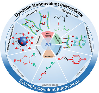
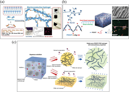
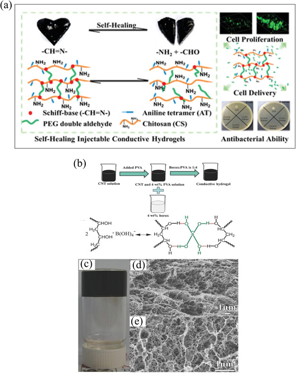
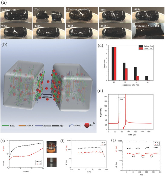
Conductive hydrogels (CHs) are emerging as a promising and well-utilized platform for 3D cell culture and tissue engineering to incorporate electron signals as biorelevant physical cues. In conventional covalently crosslinked conductive hydrogels, the network dynamics (e.g., stress relaxation, shear shining, and self-healing) required for complex cellular functions and many biomedical utilities (e.g., injection) cannot be easily realized. In contrast, dynamic conductive hydrogels (DCHs) are fabricated by dynamic and reversible crosslinks. By allowing for the breaking and reforming of the reversible linkages, DCHs can provide dynamic environments for cellular functions while maintaining matrix integrity. These dynamic materials can mimic some properties of native tissues, making them well-suited for several biotechnological and medical applications. An overview of the design, synthesis, and engineering of DCHs is presented in this review, focusing on the different dynamic crosslinking mechanisms of DCHs and their biomedical applications.

Keywords: biomedical applications; conductive hydrogels; extracellular matrix; reversible networks.


Conflict of interest statement

The authors declare no conflict of interest.

Figures

Figure 1
Figure 1
Different reversible covalent bonds and noncovalent interactions used in assembling dynamic conductive hydrogels (DCHs).
Figure 2
Figure 2
a) PEG–peptide and PEDOT:PSS interaction form a DCH via a self‐assembly process. The DCHs are self‐healable and injectable due to the reversible non‐covalent interactions and are utilized as a 3D cell culture matrix with high biocompatibility in vitro. Adapted with permission.[ 71 ] Copyright 2018, American Chemical Society. b) Pβ‐CD and Ad‐modified conductive polymer (PEDOT:S‐Alg‐Ad) formed a DCH. Adapted with permission.[ 76 ] Copyright 2019, American Chemical Society. c) The process to prepare the pure a PEDOT:PSS hydrogel with DMSO as an additive. Adapted with permission.[ 79 ] Copyright 2019, Springer Nature.
Figure 3
Figure 3
a) DCHs based on Schiff bases. DCHs were demonstrated with cell delivery and antibacterial applications. Adapted with permission.[ 98 ] Copyright 2016, American Chemical Society. b) The dynamic borate ester bond crosslinking reaction formed DCHs between PVA, SWCNT, and borate ester. c) Picture of SWCNT‐containing DCHs. d,e) Scanning electron microscopy (SEM) images of the SWCNT DCHs. Reproduced with permission.[ 108 ] Copyright 2017, Wiley‐VCH GmbH.
Figure 4
Figure 4
A double‐network DCH based on ionic interactions and radical polymerization. a) Picture of the self‐healing property of the double‐network DCH. b) Schematic demonstration of the self‐healing capability of the double‐network DCH. c) Capability of mechanical recovery after cutting and self‐healing. d) Capability of electrical recovery after cutting, showing that the electrical recovery efficiency is 96% within 1 min. e–g) Rheological testing of the double‐network DCH.Frequency sweep (e) and amplitude (f) sweep test. G′ and G″ of the double‐network DCH with cycle oscillation force (g). Adapted with permission.[ 129 ] Copyright 2017, Wiley‐VCH GmbH.
Figure 5
Figure 5
a‐i) Schematic illustration of DCH (eCA‐gel) preparation. ii) Representative live‐dead staining images of neonatal rat cardiac cells cultured on 2D at 48 h. iii) Quantitative analysis of cell viability. iv) Representative live‐dead staining images of neonatal rat cardiac cells in 3D cultured at 48 h. v) Cell viability of live‐dead staining (n = 3). Scale bars: 50 µm. Adapted with permission.[ 74 ] Copyright 2017, Wiley‐VCH. b‐i) The formation of DCH with the injection of pregel into PBS solution and a schematic illustration of the interaction mechanism. ii) Image of the electrical stimulation device for cell culture and a schematic illustration of cell culture under electrical stimulation. iii) The bundled axons in the DCH with 3D cell culture. The asterisk‐marked area in (iii) was investigated by analyzing iv) confocal images with high magnification of single optic section. v) The reconstructed axons and myelinated segments were colored green and red,respectively. vi) mRNA expression of MBP and c‐Jun genes. Adapted with permission.[ 132 ] Copyright 2020, American Chemical Society.
Figure 6
Figure 6
a‐i) The fabrication process of the adhesive PDA‐GO‐PAM based DCH. ii) The DCH acted as an adhesive electrode to detect electromyography signals. Adapted with permission.[ 138 ] Copyright 2017, Wiley‐VCH GmbH. b) The dual cross‐linked DCH adhesive. i) The preparation process of the DCHs. ii) Image of the lap‐shear experimental of DCH and adhesion mechanism. iii) Adhesion of the DCH on muscle tissue. The hydrogel remains adhered to the muscle tissue in water. Adapted with permission.[ 139 ] Copyright 2019, Wiley‐VCH GmbH.
Figure 7
Figure 7
DCH‐based implantable hydrogel bioelectronics. a‐i) Schematic illustration of the formation of DCH and its application as heart patches. ii) Representative images for nontreatment control, MI, nonconductive (HPAE/Geln), and DCH (HPAE–Py/Geln) groups after 4 weeks. iii) Representative ECGs of different groups. iv) Quantification of the QRS interval of different groups. Adapted with permission.[ 159 ] Copyright 2018, Wiley‐VCH GmbH. b‐i) Illustration of the fabrication of a DCH through crosslinking by mixing tannic acid, pyrrole, and Fe3+. ii) DCH induced endogenous neurogenesis in vivo. iii) Schematic demonstration of a DCH that was transimplanted into the spinal cord hemisection gap. iv) Quantification of the average cystic cavity area of animals with SCI and different hydrogel treatments. Adapted with permission.[ 160 ] Copyright 2018, American Chemical Society.

References

    1. Mouw J. K., Ou G., Weaver V. M., Nat. Rev. Mol. Cell Biol. 2014, 15, 771. - PMC - PubMed
    1. Theocharis A. D., Skandalis S. S., Gialeli C., Karamanos N. K., Adv. Drug Delivery Rev. 2016, 97, 4. - PubMed
    1. Frantz C., Stewart K. M., Weaver V. M., J. Cell Sci. 2010, 123, 4195. - PMC - PubMed
    1. Raeburn J., Cardoso A. Z., Adams D. J., Chem. Soc. Rev. 2013, 42, 5143. - PubMed
    1. Kharkar P. M., Kiick K. L., Kloxin A. M., Chem. Soc. Rev. 2013, 42, 7335. - PMC - PubMed

Publication types

LinkOut - more resources