. 2021 Aug 15:1765:147507.
doi: 10.1016/j.brainres.2021.147507. Epub 2021 Apr 28.

Effect of hemopexin treatment on outcome after intracerebral hemorrhage in mice

Affiliations

Effect of hemopexin treatment on outcome after intracerebral hemorrhage in mice

Jing Chen-Roetling et al. Brain Res. .

Abstract

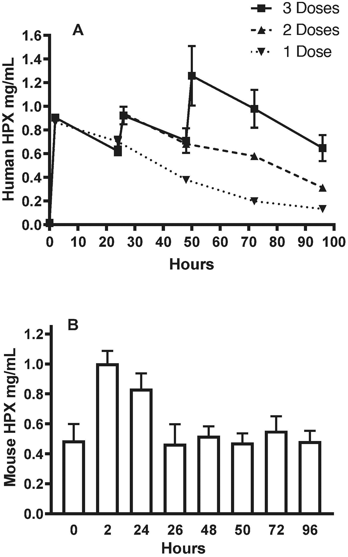
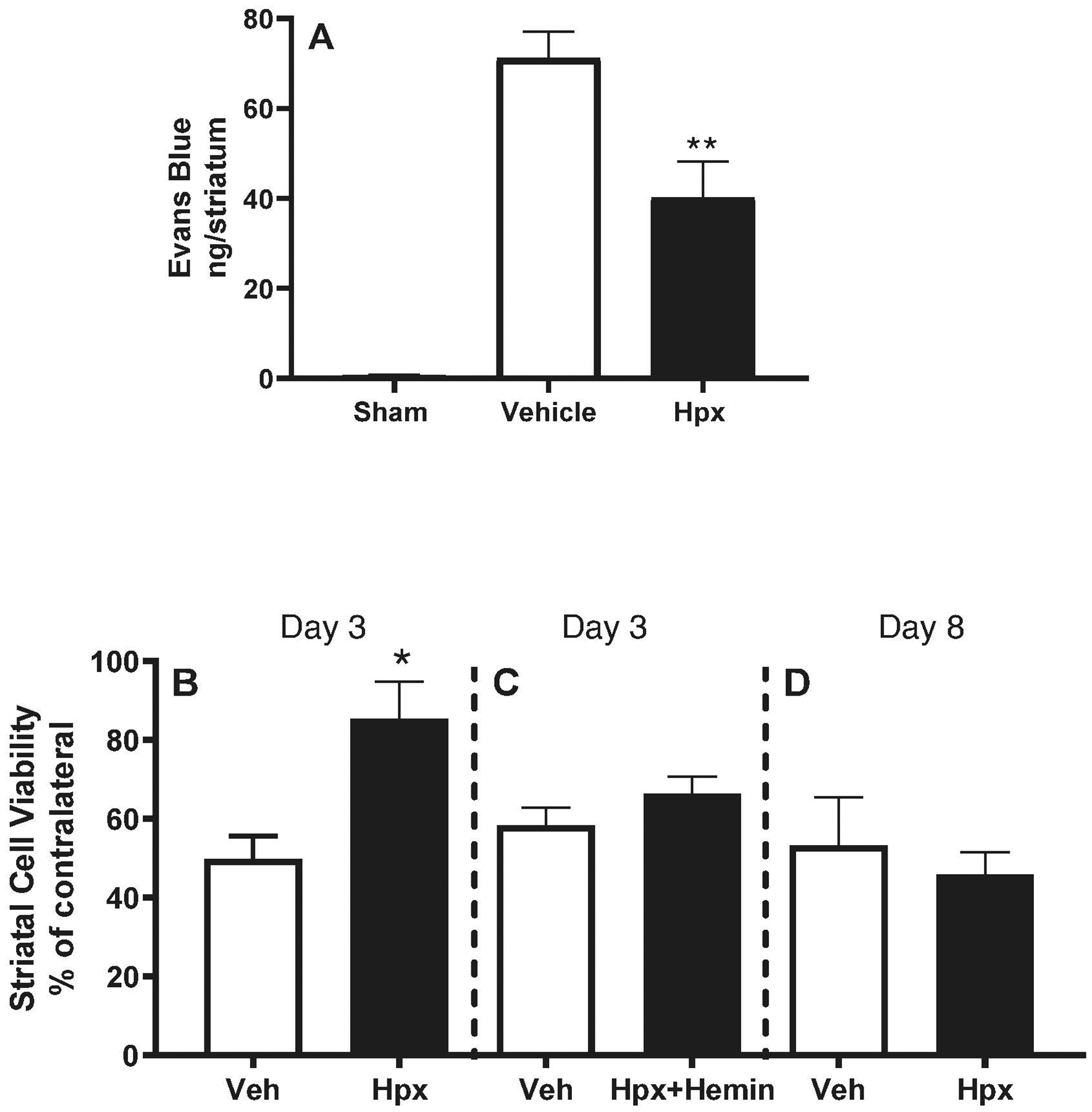
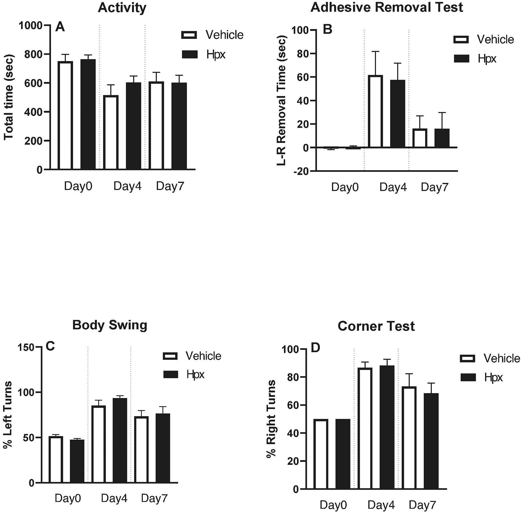
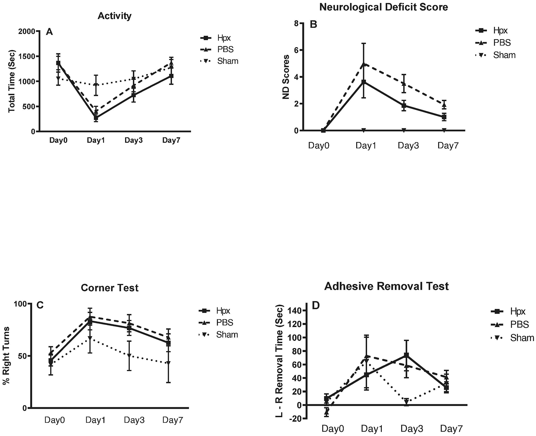
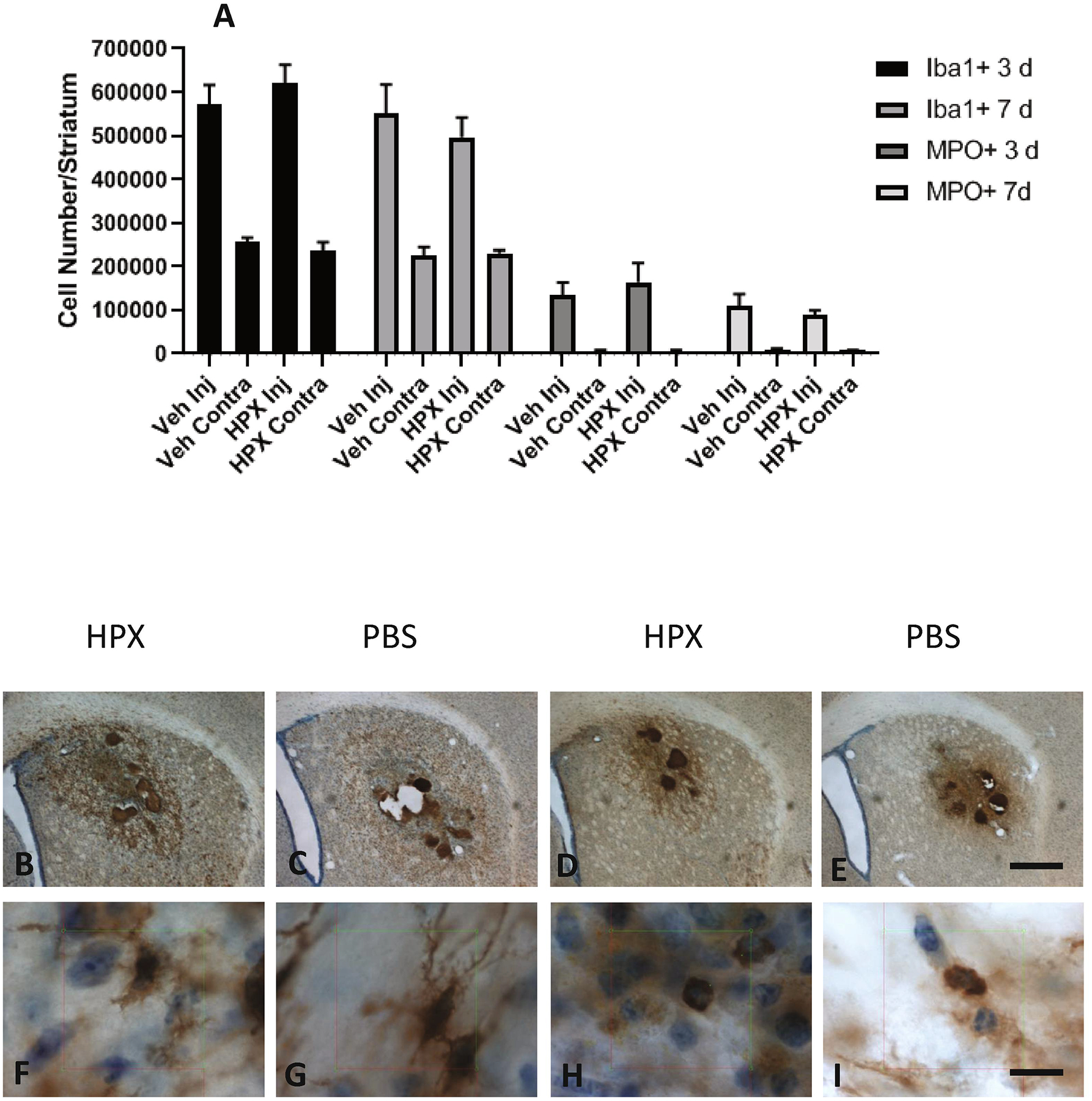
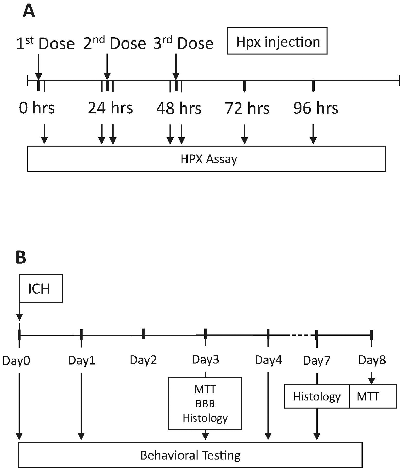
Heme release from hemoglobin may contribute to secondary injury after intracerebral hemorrhage (ICH). The primary endogenous defense against heme toxicity is hemopexin, a 57 kDa glycoprotein that is depleted in the CNS after hemorrhagic stroke. We hypothesized that systemic administration of exogenous hemopexin would reduce perihematomal injury and improve outcome after experimental ICH. Intraperitoneal treatment with purified human plasma hemopexin beginning 2 h after striatal ICH induction and repeated daily for the following two days reduced blood-brain barrier disruption and cell death at 3 days. However, it had no effect on neurological deficits at 4 or 7 days or striatal cell viability at 8 days. Continuous daily hemopexin administration had no effect on striatal heme content at 3 or 7 days, and did not attenuate neurological deficits, inflammatory cell infiltration, or perihematomal cell viability at 8 days. These results suggest that systemic hemopexin treatment reduces early injury after ICH, but this effect is not sustained, perhaps due to an imbalance between striatal tissue heme and hemopexin content at later time points. Future studies should investigate its effect when administered by methods that more efficiently target CNS delivery.

Keywords: Heme; Hemoglobin toxicity; Iron; Stroke; Stroke models; Subararachnoid hemorrhage.


Conflict of interest statement

Declarations of interest: None

Figures

Figure 1.
Figure 1.
Effect of human hemopexin (Hpx) injections on serum Hpx concentrations. A) Mice were injected with 70 mg/kg human Hpx i.p. as a single dose (0 hour), or with two or three doses separated by 24-hour intervals (0 hour, 24 hours, 48 hours). Blood was collected from a tail vein for human Hpx immunoassay at baseline and at 2h, 24h (before second dose), 26h, 48h (before third dose), 50h, 72h and 96h. B) Same serum samples as in A were tested for mouse Hpx (mean ± S.E.M., 4–8 mice/condition).
Figure 2.
Figure 2.
Early protective effect of hemopexin therapy. Right striatal ICH was induced in mice with collagenase, followed 2 hours later by 70 mg/kg Hpx i.p., 70 mg/kg Hpx with equimolar hemin, or PBS vehicle (Veh); treatments were repeated 24 and 48 hours later. A) Right striatal blood-brain barrier permeability was quantified on Day 3 by Evans blue assay (mean ± S.E.M, n=7/condition for treatment groups and 5/condition for sham); B) Right striatal cell viability at 3 days was quantified by MTT conversion for foramazan, normalized to the contralateral striatum (=100, n = 5/condition); C) Mice were treated with vehicle or heme-saturated Hpx (n = 14/condition); D) Mice were treated with 70 mg/kg Hpx for 3 doses as in A; cell viability was assessed on Day 8 (n = 10/condition). *P < 0.05, **P < 0.01 v. corresponding vehicle condition.
Figure 3.
Figure 3.
Hemopexin treatment (3 doses) had no effect on behavioral outcome. Neurological deficits were assessed at 4 and 7 days after collagenase-induced ICH by: (A) digital quantification of motor activity during 30 minute video while in standard mouse cage; (B) adhesive removal test; (C) elevated body swing test; (D) corner test. Bars represent mean ± S.E.M., n = 10/condition.
Figure 4.
Figure 4.
Effect of daily hemopexin treatment (total 7 doses) on behavioral outcome. ICH was induced by right striatal autologous blood injection, followed at 2 hours by 70 mg/kg Hpx or vehicle i.p., repeated at 24 hour intervals for 7 days. Neurological deficits were assessed at indicated time points by: (A) home cage motor activity quantification (60 minute video); (B) neurological deficit score; (C) corner test; (D) adhesive removal test. Each point represents mean ± S.E.M., 7–17 mice/condition for treatment groups and 5–8 mice/condition for sham groups.
Figure 5.
Figure 5.
Hemopexin treatment has no effect on striatal inflammatory cell numbers after ICH. A) Number of Iba1 and myeloperoxidase positive cells in hemorrhagic (Injected, Inj) and contralateral (Contra) striata at indicated time points after hemorrhage induction in mice (6–8/condition) treated with hemopexin 70 mg/kg or vehicle. Hemopexin treatment began at 2 hours and was repeated at 24 hour intervals until completion of the experiment. B-I) Representative photos captured from stereology counting session of sections from mice at 7 days after striatal blood injection, immunostained with anti-Iba1 (B,C,F,G)or myeloperoxidase (MPO, D,E,H,I). Scale bars 500 μm B-E, 10 μm F-I).
Figure 6.
Figure 6.
Timeline of experiments testing: A) effect of Hpx treatment on human and mouse serum Hpx levels; B) effect of Hpx treatment on striatal cell viability (MTT assay), blood-brain barrier (BBB) disruption (Evans blue assay), histology and behavioral deficits after ICH.

References

    1. Bai Q, Liu J, Wang G, 2020. Ferroptosis, a Regulated Neuronal Cell Death Type After Intracerebral Hemorrhage. Front Cell Neurosci. 14, 591874. - PMC - PubMed
    1. Bakker WW, Borghuis T, Harmsen MC, van den Berg A, Kema IP, Niezen KE, Kapojos JJ, 2005. Protease activity of plasma hemopexin. Kidney International. 68, 603–10. - PubMed
    1. Bakker WW, Melgert BN, Faas MM, 2010. Hemopexin: anti-inflammatory, pro-inflammatory, or both? Journal of Leukocyte Biology. 87, 1–2; author reply 3, 5. - PubMed
    1. Belcher JD, Chen C, Nguyen J, Abdulla F, Zhang P, Nguyen H, Nguyen P, Killeen T, Miescher SM, Brinkman N, Nath KA, Steer CJ, Vercellotti GM, 2018. Haptoglobin and hemopexin inhibit vaso-occlusion and inflammation in murine sickle cell disease: Role of heme oxygenase-1 induction. PLoS One. 13, e0196455. - PMC - PubMed
    1. Chen-Roetling J, Lu X, Regan KA, Regan RF, 2013. A rapid fluorescent method to quantify neuronal loss after experimental intracerebral hemorrhage. J Neurosci Methods. 216, 128–36. - PMC - PubMed

Publication types

LinkOut - more resources