. 2021 Jan-Jun:296:100715.
doi: 10.1016/j.jbc.2021.100715. Epub 2021 Apr 28.

Regulation of tau internalization, degradation, and seeding by LRP1 reveals multiple pathways for tau catabolism

Affiliations

Regulation of tau internalization, degradation, and seeding by LRP1 reveals multiple pathways for tau catabolism

Joanna M Cooper et al. J Biol Chem. 2021 Jan-Jun.

Abstract

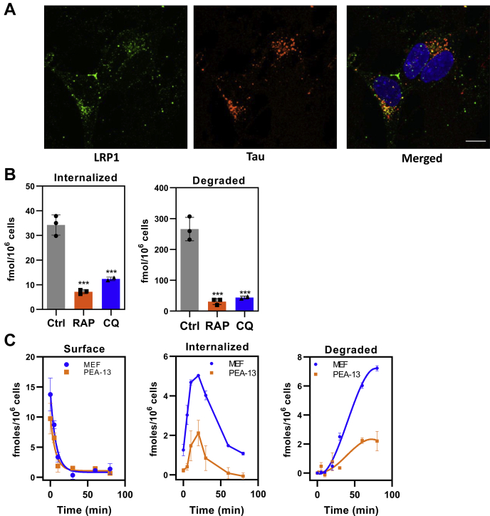
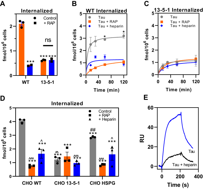
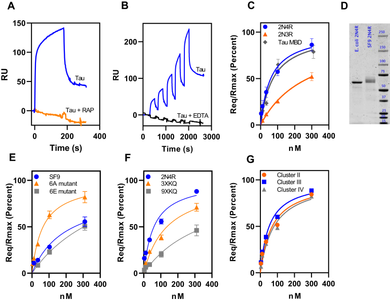
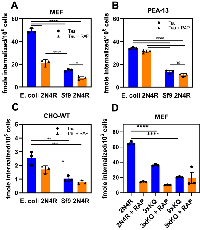
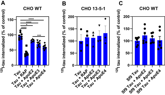
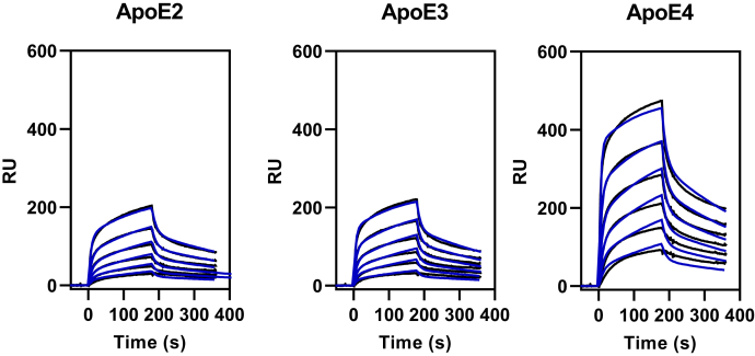
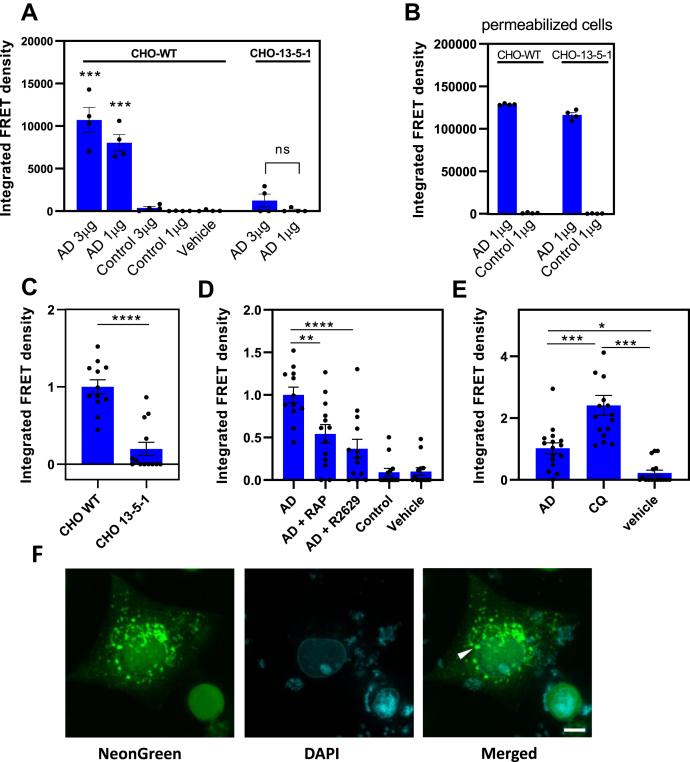
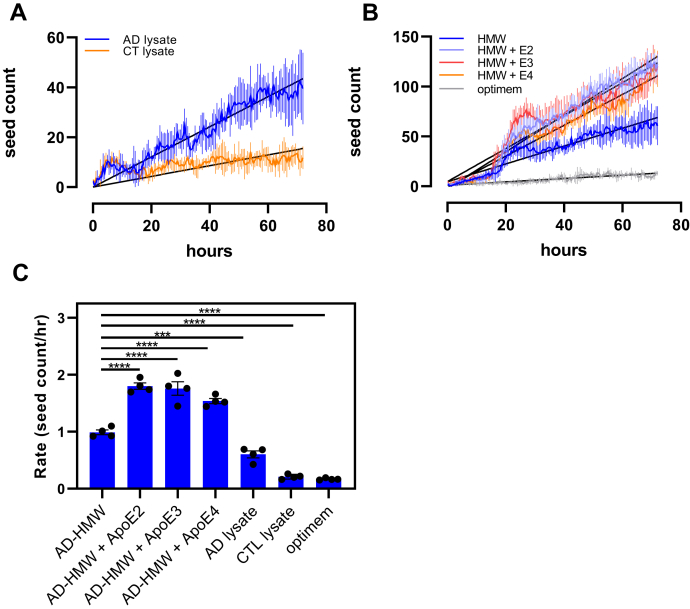
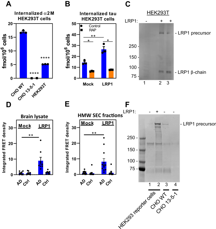
In Alzheimer's disease (AD), pathological forms of tau are transferred from cell to cell and "seed" aggregation of cytoplasmic tau. Phosphorylation of tau plays a key role in neurodegenerative tauopathies. In addition, apolipoprotein E (apoE), a major component of lipoproteins in the brain, is a genetic risk determinant for AD. The identification of the apoE receptor, low-density lipoprotein receptor-related protein 1 (LRP1), as an endocytic receptor for tau raises several questions about the role of LRP1 in tauopathies: is internalized tau, like other LRP1 ligands, delivered to lysosomes for degradation, and does LRP1 internalize pathological tau leading to cytosolic seeding? We found that LRP1 rapidly internalizes 125I-labeled tau, which is then efficiently degraded in lysosomal compartments. Surface plasmon resonance experiments confirm high affinity binding of tau and the tau microtubule-binding domain to LRP1. Interestingly, phosphorylated forms of recombinant tau bind weakly to LRP1 and are less efficiently internalized by LRP1. LRP1-mediated uptake of tau is inhibited by apoE, with the apoE4 isoform being the most potent inhibitor, likely because of its higher affinity for LRP1. Employing post-translationally-modified tau derived from brain lysates of human AD brain tissue, we found that LRP1-expressing cells, but not LRP1-deficient cells, promote cytosolic tau seeding in a process enhanced by apoE. These studies identify LRP1 as an endocytic receptor that binds and processes monomeric forms of tau leading to its degradation and promotes seeding by pathological forms of tau. The balance of these processes may be fundamental to the spread of neuropathology across the brain in AD.

Keywords: Alzheimer' s disease; Apolipoprotien E; LRP1; catabolism; lipoprotein receptors; neurofibrillary tangles; neurons; surface plasmon resonance; tau; tau spreading.


Conflict of interest statement

Conflict of interest The authors declare that they have no conflicts of interest with the contents of this article.

Figures

Figure 1
Figure 1
Tau is efficiently degraded following LRP1-mediated internalization.A, human neuroblastoma cells (SH-SY5Y) were incubated with monoclonal antibody 5A6 conjugated with Alexa Fluor 488 (green) to label the endocytic pool of LRP1, followed by 20 nM tau conjugated with Alexa Fluor 594 (red). The cells were then fixed and imaged. Colocalization of functional LRP1 and internalized tau is displayed on merged panel (yellow). The scale bar represents 10 μm. B, WI-38 cells were incubated with 20 nM 125I-labeled tau in the absence or the presence of 1 μM RAP for 2 h at 37 °C, and the steady-state levels internalized (right panel) and degraded (left panel) quantified. Internalization and degradation were also measured in the presence or the absence of 100 μM chloroquine (CQ) (shown are means ± SEM; one-way ANOVA followed by Tukey's multiple comparisons test) (∗∗∗p < 0.001 compared with control, n = 3). C, MEF cells or LRP1-deficient PEA-13 cells were incubated with 20 nM 125I-labeled tau at 4 °C for 2 h in the presence or the absence of 1 μM RAP, then the media were replaced with warm assay media, and cells were incubated at 37 °C for specified times. The amount of RAP-sensitive surface bound, internalized, and degraded 125I-labeled tau was quantified (shown are means ± SEM, n = 3). LRP1, low-density lipoprotein receptor–related protein 1; MEF, mouse embryonic fibroblast; RAP, receptor-associated protein.
Figure 2
Figure 2
Uptake of tau in LRP1-deficient CHO cells confirms the existence of additional receptors for tau uptake.A, steady-state levels of 125I-labeled tau (20 nM) internalized in WT or LRP1-deficient 13-5-1 CHO cells when incubated in the absence or the presence of 1 μM RAP for 2 h at 37 °C. B and C, time course for internalization of 125I-labeled tau (20 nM) in CHO WT (B) and CHO 13-5-1 (C) cells in the presence or the absence of RAP (1 mM) or heparin (20 mg/ml). D, WT, 13-5-1 and HSPG-deficient (CHO HSPG) CHO cells were incubated with 20 nM 125I-labeled tau in the absence or the presence of RAP (1 mM) or heparin (20 mg/ml) at 37 °C for 2 h, and internalized tau was measured. E, SPR analysis of 10 nM tau binding to LRP1 in the absence or the presence of 20 mg/ml heparin. AD, means ± SEM; two-way ANOVA followed by Sidak's multiple comparisons test, (A) ∗∗∗p < 0.0001 compared with WT control, n = 3; (B and C) ∗p < 0.0001 comparison of tau versus tau + RAP, n = 3; (D) significance reported compared with ∗CHO WT, #CHO 13-5-1, or ˆCHO HSPG (one symbol p < 0.03; two symbols p < 0.007; and three symbols p < 0.0001). CHO, Chinese hamster ovary; HSPG, heparan sulfate proteoglycan; LRP1, low-density lipoprotein receptor–related protein 1; RAP, receptor-associated protein; SPR, surface plasmon resonance.
Figure 3
Figure 3
Phosphorylated forms of tau bind weakly to LRP1.A, inhibition of tau binding to LRP1 by excess RAP as assessed by coinjection experiment. B, single-cycle kinetic experiment quantifying binding of monomeric tau (3.8, 11.5, 34.4, 103.3, and 310 nM) to LRP1 in the presence of Ca2+ (blue line) or EDTA (black line). C, binding of tau isoforms 2N4R, 2N3R, and tau MBD to LRP1 assessed by SPR equilibrium analysis. D, about 1 μg of recombinant tau produced in Escherichia coli or SF9 cells was ran on a 4 to 12% gel and stained with colloidal Coomassie. Image was captured using Licor. Quantification of bands reveals a signal of 2270 for E. coli tau and 2280 for SF9 tau. E, the binding of tau produced by Sf9 cells along with two mutant forms of tau to full-length human LRP1 was measured by SPR; 6A (T181, S199, S202, S396, S400, and S404 are all converted to alanine) and 6E, in which all these residues are converted to glutamic acid. F, binding of mutant forms of tau to LRP1: 3XKQ in which lysine residues 311, 317, and 321 were converted to glutamine residues and 9XKQ tau in which lysine residues 311, 217, 321, 340, 343, 347, 353, 369, and 375 are all converted to glutamine residues. G, binding of monomeric tau to LRP1 clusters II, III, or IV by SPR equilibrium analysis. For all experiments, n = 3 (biological replicates), (A and B) show representative data, (C, E, and F) show means ± SEM. LRP1, low-density lipoprotein receptor–related protein 1; MBD, microtubule-binding domain; RAP, receptor-associated protein; SPR, surface plasmon resonance.
Figure 4
Figure 4
Hyperphosphorylated forms of tau are not efficiently internalized by LRP1. Internalization of 20 nM 125I-labeled WT tau or Sf9-produced tau in MEF cells (A) LRP1-deficient PEA-13 cells (B) or CHO-WT cells (C) in the presence or the absence of RAP; D, internalization of 20 nM of 125I-labeled WT tau, 3XKQ, or 9XKQ mutants. (Two-way ANOVA, Sidak's multiple comparisons test; ∗∗∗∗p < 0.0001, ∗∗∗p < 0.0007, ∗∗p < 0.002, ∗p < 0.04, n = 3). CHO, Chinese hamster ovary; LRP1, low-density lipoprotein receptor–related protein 1; MEF, mouse embryonic fibroblast; RAP, receptor-associated protein.
Figure 5
Figure 5
ApoE reduces LRP1-mediated tau internalization but not hyperphosphorylated tau. WT CHO (A and C) or LRP1-deficient CHO 13-5-1 cells (B) were cultured overnight in the presence of 10 μg/ml apoE2, apoE3, or apoE4. The cells were then incubated with 20 nM 125I-tau from Escherichia coli (A and B) or 100 nM of hyperphosphorylated 125I-tau isolated from SF9 cells (C) in the absence or the presence of 10 μg/ml apoE isoforms at 37 °C for 2 h. The amounts of internalized 125I-tau were quantified. Statistical analysis was performed using one-way ANOVA and Tukey's post hoc test (∗p < 0.05, ∗∗∗p < 0.0006, and ∗∗∗∗p < 0.0001). ApoE, apolipoprotein E; CHO, Chinese hamster ovary; LRP1, low-density lipoprotein receptor–related protein 1.
Figure 6
Figure 6
SPR analysis of apoE isoforms binding to LRP1. Increasing concentrations of apoE2, apoE3, or apoE4 (52, 104, 208, 417, 834, and 1668 nM) were injected over the LRP1-coupled chip. Fits of the experimental data to a bivalent binding model are shown as blue lines. The data shown are a representative experiment from six independent experiments that were performed. apoE, apolipoprotein E; LRP1, low-density lipoprotein receptor–related protein 1; SPR, surface plasmon resonance.
Figure 7
Figure 7
LRP1 mediates tau seeding.A, CHO WT and 13-5-1 cells were transfected with pcDNA3 plasmid containing a construct that encoded residues 344 to 378 of human P301L mutant tau fused to either mTurquoise2 or mNeonGreen and then incubated with 1 to 3 μg of human brain homogenate from a patient with AD or from a healthy control for 24 h. B, as a positive control, 1% Lipofectamine 2000 was added to the wells. A and B, tau seeding was quantified by multiplying the percent of FRET-positive cells by the median florescence intensity of those cells. Each condition was performed in at least quadruplicate, and data were analyzed using FlowJo software. Means ± SEM, one-way ANOVA followed by Tukey's multiple comparisons test ∗∗∗p < 0.0001 compared with vehicle control. C, transfected CHO WT and 13-5-1 cells were incubated with HMW-SEC fractions from human brain of a patient with AD, shown are means ± SEM, t test; n = 12, ∗∗∗∗p < 0.0001. D, CHO WT cells transfected with the tau FRET reporter system were incubated with HMW-SEC fractions from human brain of a patient with AD in the presence or the absence of 1 μM RAP or anti-LRP1 IgG (R2629). E, CHO WT cells transfected with the tau FRET reporter system were incubated with HMW-SEC fractions from human brain of a patient with AD in the absence or the presence of 100 μM chloroquine (CQ) (means ± SEM, one-way ANOVA followed by Tukey's multiple comparisons test ∗∗∗p < 0.0001, ∗p < 0.03). F, representative images of foci of aggregated FRET reporter in CHO WT cells incubated with HMW-SEC tau, showing native florescence of mNeonGreen. The scale bar represents 10 μm. AD, Alzheimer's disease; CHO, Chinese hamster ovary; HMW-SEC, high–molecular weight seeding-competent; LRP1, low-density lipoprotein receptor–related protein 1; RAP, receptor-associated protein.
Figure 8
Figure 8
ApoE isoforms increase the rate of seeding in stably transfected CHO WT cells. CHO WT cells were stably transfected with pcDNA3 plasmid containing a construct that encodes residues 344 to 378 of human P301L mutant tau fused to either mTurquoise2 or mNeonGreen. A, the cells were incubated with lysates from patients with AD (AD) or age-matched controls (CTL), and seeding was monitored as a function of time. B, cells incubated with HMW-SEC fractions from human brain AD patient in the absence or the presence of apoE2, apoE3, or apoE4. Linear regression analysis was performed (black lines) to estimate the rate of seeding. (HMW, r2 = 0.9274; HMW + ApoE2, r2 = 0.9688; HMW + ApoE3, r2 = 0.9215; HMW + AppE4, r2 = 0/9574). C, rates of seeding are plotted for each treatment. Shown are means ± SEM. n = 4, one-way ANOVA followed by Tukey's multiple comparisons test to AD-HMW. ∗∗∗∗p < 0.0001 and ∗∗∗p < 0.0009. AD, Alzheimer's disease; ApoE, apolipoprotein E; CHO, Chinese hamster ovary; HMW-SEC, high–molecular weight seeding-competent.
Figure 9
Figure 9
LRP1 mediates tau seeding in HEK293T cells.A, CHO WT, 13-5-1, and HEK293T cells were incubated with 5 nM 125I-labeled a2M∗ for 2 h, and the amount internalized quantified. B, HEK293T cells were mock transfected or transfected with LRP1 plasmid. About 24 h after transfection, cells were incubated with 125I-tau (20 nM) in the presence or the absence of 1 μM RAP for 2 h, and internalized tau was quantified (n = 3, technical replicates). C, immunoblot of cells used in (B) with anti-LRP1 IgG. HEK293T FRET reporter cells were transfected with LRP1 and then incubated with (D) human brain homogenate from an Alzheimer's patient (n = 8, two biological replicates containing four technical replicates each) or (E) with HMW-SEC fractions from AD patient brain (n = 12, three biological replicates containing four technical replicates each). F, immunoblot of HEK283T reporter cells along with CHO WT and CHO LRP1-deficient CHO 13-5-1 cells (as controls). ((A) Means ± SEM, one-way ANOVA or (B, D, and E) two-way ANOVA followed by Tukey's multiple comparisons test. ∗p < 0.05, ∗∗p < 0.01, and ∗∗∗∗p < 0.0001). AD, Alzheimer's disease; CHO, Chinese hamster ovary; HEK293T, human embryonic kidney 293T; HMW-SEC, high–molecular weight seeding-competent; LRP1, low-density lipoprotein receptor–related protein 1; RAP, receptor-associated protein.
Figure 10
Figure 10
Surface-accessible lysine residues available for LRP1 binding on tau protofilament from Alzheimer's disease.Ribbon diagram of tau filament core (Protein Data Bank: 5O3L) (70) showing accessible surface areas (ASAs) for lysine residues available for interacting with LRP1 (ASA > 0.5). ASA was calculated from the coordinates in Protein Data Bank 5O3L using Volume, Area, Dihedral Angle Reporter, version 1.8 (71). Distances between the a-carbon of lysine residues were determined using PyMOL software. ∗Lysine resides mutated in 3XKQ tau mutant; ∗∗additional lysine residues mutated in 9XKQ mutant. LRP1, low-density lipoprotein receptor–related protein 1.
None

Similar articles

Cited by

References

    1. Hyman B.T., Van Hoesen G.W., Damasio A.R., Clifford L. Alzheimer’s disease: Cell-specific pathology isolates the hippocampal formation. Science. 1984;225:1168–1170. - PubMed
    1. Serrano-Pozo A., Qian J., Monsell S.E., Frosch M.P., Betensky R.A., Hyman B.T. Examination of the clinicopathologic continuum of Alzheimer disease in the autopsy cohort of the National Alzheimer Coordinating Center. J. Neuropathol. Exp. Neurol. 2013;72:1182–1192. - PMC - PubMed
    1. Braak H., Braak E. Neuropathological stageing of Alzheimer-related changes. Acta Neuropathol. 1991;82:239–259. - PubMed
    1. Polydoro M., de Calignon A., Suárez-Calvet M., Sanchez L., Kay K.R., Nicholls S.B., Roe A.D., Pitstick R., Carlson G.A., Gómez-Isla T., Spires-Jones T.L., Hyman B.T. Reversal of neurofibrillary tangles and tau-associated phenotype in the rTgTauEC model of early Alzheimer’s disease. J. Neurosci. 2013;33:13300–13311. - PMC - PubMed
    1. De Calignon A., Polydoro M., Suárez-Calvet M., William C., Adamowicz D.H., Kopeikina K.J., Pitstick R., Sahara N., Ashe K.H., Carlson G.A., Spires-Jones T.L., Hyman B.T. Propagation of tau pathology in a model of early Alzheimer’s disease. Neuron. 2012;73:685–697. - PMC - PubMed

Publication types

Substances