

Life Event Stress and Reduced Cortical Thickness in Youth at Clinical High Risk for Psychosis and Healthy Control Subjects
- PMID: 33930604
- PMCID: PMC8551305
- DOI: 10.1016/j.bpsc.2021.04.011
Life Event Stress and Reduced Cortical Thickness in Youth at Clinical High Risk for Psychosis and Healthy Control Subjects
Abstract
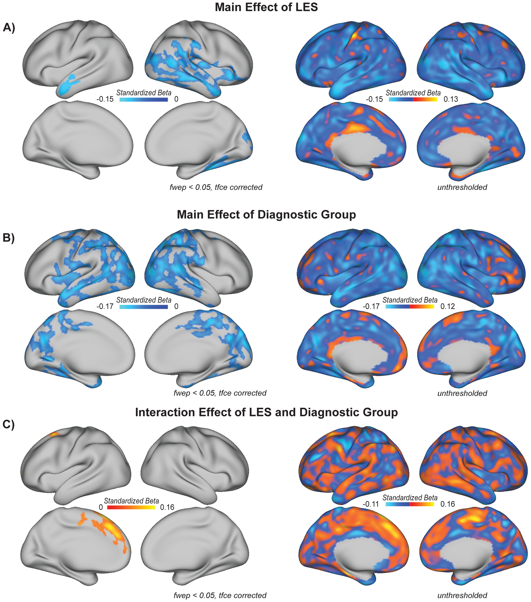
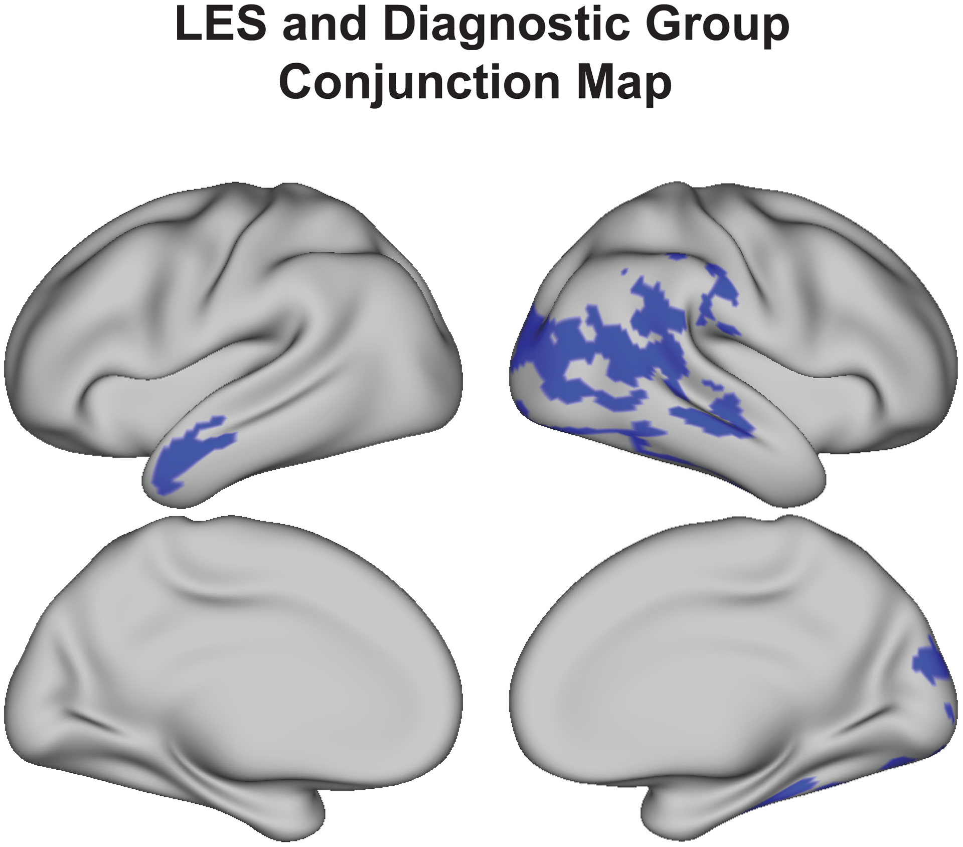
Background: A decline in cortical thickness during early life appears to be a normal neuromaturational process. Accelerated cortical thinning has been linked with conversion to psychosis among individuals at clinical high risk for psychosis (CHR-P). Previous research indicates that exposure to life event stress (LES) is associated with exaggerated cortical thinning in both healthy and clinical populations, and LES is also linked with conversion to psychosis in CHR-P. To date, there are no reports on the relationship of LES with cortical thickness in CHR-P. This study examines this relationship and whether LES is linked with cortical thinning to a greater degree in individuals at CHR-P who convert to psychosis compared with individuals at CHR-P who do not convert and healthy control subjects.
Methods: Controlling for age and gender (364 male, 262 female), this study examined associations between LES and baseline cortical thickness in 436 individuals at CHR-P (375 nonconverters and 61 converters) and 190 comparison subjects in the North American Prodrome Longitudinal Study.
Results: Findings indicate that prebaseline cumulative LES is associated with reduced baseline cortical thickness in several regions among the CHR-P and control groups. Evidence suggests that LES is a risk factor for thinner cortex to the same extent across diagnostic groups, while CHR-P status is linked with thinner cortex in select regions after accounting for LES.
Conclusions: This research provides additional evidence to support the role of LES in cortical thinning in both healthy youth and those at CHR-P. Potential underlying mechanisms of the findings and implications for future research are discussed.
Keywords: Clinical high risk; Cortical thickness; Environment; Life stress; Neuromaturation; Psychosis.
Copyright © 2021 Society of Biological Psychiatry. Published by Elsevier Inc. All rights reserved.
Conflict of interest statement
Disclosures
All authors report no biomedical financial interests or potential conflicts of interest.
Figures
Comment in
-
Advances and Future Directions in Understanding Associations Between Stressful Events and Cortical Thickness in Psychosis Risk.Biol Psychiatry Cogn Neurosci Neuroimaging. 2022 Feb;7(2):124-126. doi: 10.1016/j.bpsc.2021.11.001. Biol Psychiatry Cogn Neurosci Neuroimaging. 2022. PMID: 35131048 No abstract available.
References
-
- Norman RM, Malla AK. (1993): Stressful life events and schizophrenia: A review of the research. British Journal of Psychiatry 162: 161–166. - PubMed
-
- Walker EF, Diforio D. (1997): Schizophrenia: A neural diathesis-stress model. Psychological Review 104(4): 667. - PubMed
-
- Cotter D, Pariante CM. (2002): Stress and the progression of the developmental hypothesis of schizophrenia. British Journal of Psychiatry 181: 363–365. - PubMed
-
- Lysaker PH, LaRocco VA. (2008): The prevalence and correlates of trauma-related symptoms in schizophrenia spectrum disorder. Comprehensive Psychiatry 49: 330–334. - PubMed
Publication types
MeSH terms
Grants and funding
- UL1 TR001863/TR/NCATS NIH HHS/United States
- R01 MH076989/MH/NIMH NIH HHS/United States
- U01 MH081988/MH/NIMH NIH HHS/United States
- U01 MH066069/MH/NIMH NIH HHS/United States
- P50 MH066286/MH/NIMH NIH HHS/United States
- U01 MH066134/MH/NIMH NIH HHS/United States
- U01 MH082022/MH/NIMH NIH HHS/United States
- U01 MH081984/MH/NIMH NIH HHS/United States
- U01 MH081902/MH/NIMH NIH HHS/United States
- P50 HD103573/HD/NICHD NIH HHS/United States
- R01 MH060720/MH/NIMH NIH HHS/United States
- U01 MH081928/MH/NIMH NIH HHS/United States
- U01 MH081857/MH/NIMH NIH HHS/United States
- U01 MH082004/MH/NIMH NIH HHS/United States
- U01 MH081944/MH/NIMH NIH HHS/United States
LinkOut - more resources
Full Text Sources
Other Literature Sources
Medical
