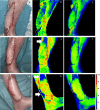
Hyperspectral Imaging (HSI) as a new diagnostic tool in free flap monitoring for soft tissue reconstruction: a proof of concept study
- PMID: 33931056
- PMCID: PMC8086299
- DOI: 10.1186/s12893-021-01232-0
Hyperspectral Imaging (HSI) as a new diagnostic tool in free flap monitoring for soft tissue reconstruction: a proof of concept study
Abstract
Objectives: Free flap surgery is an essential procedure in soft tissue reconstruction. Complications due to vascular compromise often require revision surgery or flap removal. We present hyperspectral imaging (HSI) as a new tool in flap monitoring to improve sensitivity compared to established monitoring tools.
Methods: We performed a prospective observational cohort study including 22 patients. Flap perfusion was assessed by standard clinical parameters, Doppler ultrasound, and HSI on t0 (0 h), t1 (16-28 h postoperatively), and t2 (39-77 h postoperatively). HSI records light spectra from 500 to 1000 nm and provides information on tissue morphology, composition, and physiology. These parameters contain tissue oxygenation (StO2), near-infrared perfusion- (NIR PI), tissue hemoglobin- (THI), and tissue water index (TWI).
Results: Total flap loss was seen in n = 4 and partial loss in n = 2 cases. Every patient with StO2 or NIR PI below 40 at t1 had to be revised. No single patient with StO2 or NIR PI above 40 at t1 had to be revised. Significant differences between feasable (StO2 = 49; NIR PI = 45; THI = 16; TWI = 56) and flaps with revision surgery [StO2 = 28 (p < 0.001); NIR PI = 26 (p = 0.002); THI = 56 (p = 0.002); TWI = 47 (p = 0.045)] were present in all HSI parameters at t1 and even more significant at t2 (p < 0.0001).
Conclusion: HSI provides valuable data in free flap monitoring. The technique seems to be superior to the gold standard of flap monitoring. StO2 and NIR PI deliver the most valuable data and 40 could be used as a future threshold in surgical decision making. Clinical Trial Register This study is registered at the German Clinical Trials Register (DRKS) under the registration number DRKS00020926.
Keywords: Flap surgery; Hyperspectral imaging; Imaging; Monitoring; Reconstructive surgery.
Conflict of interest statement
The authors declare that they have no competing interests.
Figures


References
-
- Jones NF, Jarrahy R, Song JI, et al. Postoperative medical complications–not microsurgical complications–negatively influence the morbidity, mortality, and true costs after microsurgical reconstruction for head and neck cancer. Plast Reconstr Surg. 2007;119:2053–2060. doi: 10.1097/01.prs.0000260591.82762.b5. - DOI - PubMed
Publication types
MeSH terms
LinkOut - more resources
Full Text Sources
Other Literature Sources
Research Materials
Miscellaneous

