A review of visualization techniques of post-mortem computed tomography data for forensic death investigations
- PMID: 33931808
- PMCID: PMC8354982
- DOI: 10.1007/s00414-021-02581-4
A review of visualization techniques of post-mortem computed tomography data for forensic death investigations
Abstract
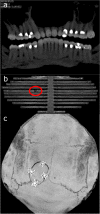
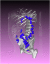
Postmortem computed tomography (PMCT) is a standard image modality used in forensic death investigations. Case- and audience-specific visualizations are vital for identifying relevant findings and communicating them appropriately. Different data types and visualization methods exist in 2D and 3D, and all of these types have specific applications. 2D visualizations are more suited for the radiological assessment of PMCT data because they allow the depiction of subtle details. 3D visualizations are better suited for creating visualizations for medical laypersons, such as state attorneys, because they maintain the anatomical context. Visualizations can be refined by using additional techniques, such as annotation or layering. Specialized methods such as 3D printing and virtual and augmented reality often require data conversion. The resulting data can also be used to combine PMCT data with other 3D data such as crime scene laser scans to create crime scene reconstructions. Knowledge of these techniques is essential for the successful handling of PMCT data in a forensic setting. In this review, we present an overview of current visualization techniques for PMCT.
Keywords: 3D printing; Postmortem computed tomography; Reporting; Segmentation; Virtual reality; Visualization.
© 2021. The Author(s).
Conflict of interest statement
The authors declare no competing interests.
Figures


Similar articles
-
An algorithm for automatically generating gas, bone and foreign body visualizations from postmortem computed tomography data.Forensic Sci Med Pathol. 2021 Jun;17(2):254-261. doi: 10.1007/s12024-021-00363-3. Epub 2021 Apr 27. Forensic Sci Med Pathol. 2021. PMID: 33905073 Free PMC article.
-
Post-mortem computed tomography (PMCT) radiological findings and assessment in advanced decomposed bodies.Radiol Med. 2019 Oct;124(10):1018-1027. doi: 10.1007/s11547-019-01052-6. Epub 2019 Jun 28. Radiol Med. 2019. PMID: 31254219
-
The role of computed tomography in post-mortem examinations.Arch Med Sadowej Kryminol. 2024;74(2):124-133. doi: 10.4467/16891716AMSIK.24.011.20340. Arch Med Sadowej Kryminol. 2024. PMID: 39470757 Review. English, Polish.
-
Layering of stomach contents in drowning cases in post-mortem computed tomography compared to forensic autopsy.Int J Legal Med. 2019 Jan;133(1):181-188. doi: 10.1007/s00414-018-1850-4. Epub 2018 Apr 24. Int J Legal Med. 2019. PMID: 29691641
-
Forensic post-mortem CT in children.Clin Radiol. 2023 Nov;78(11):839-847. doi: 10.1016/j.crad.2023.06.001. Epub 2023 Aug 14. Clin Radiol. 2023. PMID: 37827594 Review.
Cited by
-
Identification of gunshot entry wounds using hyperdense rim sign on post-mortem computed tomography.Int J Legal Med. 2025 Mar;139(2):619-626. doi: 10.1007/s00414-024-03362-5. Epub 2024 Oct 31. Int J Legal Med. 2025. PMID: 39480552
-
A Virtual, 3D Multimodal Approach to Victim and Crime Scene Reconstruction.Diagnostics (Basel). 2023 Aug 25;13(17):2764. doi: 10.3390/diagnostics13172764. Diagnostics (Basel). 2023. PMID: 37685302 Free PMC article. Review.
-
Estimating age at death by Hausdorff distance analyses of the fourth lumbar vertebral bodies using 3D postmortem CT images.Forensic Sci Med Pathol. 2024 Jun;20(2):472-479. doi: 10.1007/s12024-023-00620-7. Epub 2023 Apr 14. Forensic Sci Med Pathol. 2024. PMID: 37058209
-
The current state of forensic imaging - post mortem imaging.Int J Legal Med. 2025 May;139(3):1141-1159. doi: 10.1007/s00414-025-03461-x. Epub 2025 Mar 24. Int J Legal Med. 2025. PMID: 40126650 Free PMC article. Review.
-
Applicability and usefulness of the Declaration of Helsinki for forensic research with human cadavers and remains.Forensic Sci Med Pathol. 2023 Mar;19(1):1-7. doi: 10.1007/s12024-022-00510-4. Epub 2022 Aug 6. Forensic Sci Med Pathol. 2023. PMID: 35932421 Free PMC article.
References
-
- Carew RM, Errickson D. Imaging in forensic science: five years on. J Forensic Radiol Imaging. 2019;16:24–33. doi: 10.1016/j.jofri.2019.01.002. - DOI
-
- Ebert LC, Flach P, Schweitzer W, et al. Forensic 3D surface documentation at the Institute of forensic medicine in Zurich – workflow and communication pipeline. J Forensic Radiol Imaging. 2016;5:1–7. doi: 10.1016/j.jofri.2015.11.007. - DOI
Publication types
MeSH terms
LinkOut - more resources
Full Text Sources
Other Literature Sources
Medical

