. 2021 Aug;30(8):1577-1593.
doi: 10.1002/pro.4091. Epub 2021 May 11.

Epitope mapping of novel monoclonal antibodies to human angiotensin I-converting enzyme

Affiliations

Epitope mapping of novel monoclonal antibodies to human angiotensin I-converting enzyme

Isolda A Popova et al. Protein Sci. 2021 Aug.

Abstract

Angiotensin I-converting enzyme (ACE, CD143) plays a crucial role in blood pressure regulation, vascular remodeling, and immunity. A wide spectrum of mAbs to different epitopes on the N and C domains of human ACE have been generated and used to study different aspects of ACE biology, including establishing a novel approach-conformational fingerprinting. Here we characterized a novel set of 14 mAbs, developed against human seminal fluid ACE. The epitopes for these novel mAbs were defined using recombinant ACE constructs with truncated N and C domains, species cross-reactivity, ACE mutagenesis, and competition with the previously mapped anti-ACE mAbs. Nine mAbs recognized regions on the N domain, and 5 mAbs-on the C domain of ACE. The epitopes for most of these novel mAbs partially overlap with epitopes mapped onto ACE by the previously generated mAbs, whereas mAb 8H1 recognized yet unmapped region on the C domain where three ACE mutations associated with Alzheimer's disease are localized and is a marker for ACE mutation T877M. mAb 2H4 could be considered as a specific marker for ACE in dendritic cells. This novel set of mAbs can identify even subtle changes in human ACE conformation caused by tissue-specific glycosylation of ACE or mutations, and can detect human somatic and testicular ACE in biological fluids and tissues. Furthermore, the high reactivity of these novel mAbs provides an opportunity to study changes in the pattern of ACE expression or glycosylation in different tissues, cells, and diseases, such as sarcoidosis and Alzheimer's disease.

Keywords: ACE mutations; Alzheimer's disease; CD143; angiotensin I-converting enzyme; conformational fingerprinting; dendritic cells; glycosylation; prostate; sarcoidosis.


Conflict of interest statement

Dr. Garcia is Founder and CEO of Aqualung Therapeutics Corp. (Tucson, AZ).

Figures

FIGURE 1
FIGURE 1
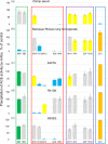
ACE activity precipitation by mAbs to ACE. (a) Purified ACE from human seminal fluid (ACE SF), adjusted to a final enzymatic activity of 5–10 mU/ml using ZPHL as a substrate, was used as a source of catalytically active ACE and incubated in a microtiter plate coated with mAbs via a goat‐anti‐mouse IgG bridge. Precipitated ACE activity was quantified by spectrofluorometric assay with ZPHL. (b) Purified ACE from human lung homogenate and ACE SF were precipitated with mAbs as in A, and presented as ACE SF/ACE lung binding ratio. Lung and SF ACE activities were equilibrated using ZPHL as a substrate prior to precipitation. Precipitation of ACE activity by mAb 9B9 in this particular graph was normalized by ACE activity in both preparations –in order to validate further normalization of ACE precipitation by other mAbs via normalization with mAb 9B9. (c) Pooled human serum from healthy blood donors (Interstate Blood Bank, Inc, Memphis, TN) diluted 1/5 with PBS and equilibrated to ACE activity in ACE SF (with ZPHL as a substrate) was precipitated by mAbs as in (a), and presented as the human serum ACE/ACE SF ratio. (d) ACE activity precipitation by tested mAbs was quantified (as in A) in the culture medium of CHO cells expressing human recombinant truncated N‐ and C‐domain constructs as well as soluble somatic ACE without the transmembrane anchor, denoted WTΔ,, equilibrated to ACE activity (with ZPHL as a substrate) and presented as N‐ or C‐domain/somatic ACE ratio. Data are presented as the mean percentage of ACE activity precipitated by the indicated mAbs, determined in three independent experiments (which did not differ by more than 10%) relative to that precipitated by mAb 9B9., mAb 9B9 is indicated by a green bar, orange bars indicate an increase of ACE precipitation of more than 20% over 100%, brown bars indicate an increase of more than 50%, red bars indicate an increase of more than two‐fold, yellow bars indicate a decrease in ACE precipitation of more than 20% over 100%, blue bars indicate a decrease of more than 50%
FIGURE 2
FIGURE 2
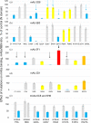
ACE activity precipitation by mAbs to the N domain of ACE. Chimpanzee serum (a) or macaque rhesus lung homogenate (b) were used as sources of catalytically active ACEs. The culture medium of CHO cells expressing the mutated forms of the truncated N domain was used as a source of N domain mutants (c–e). Human serum, lung homogenate, and culture medium of CHO cells expressing truncated N domain were used as controls. mAbs 9B9 and i2H5/1G12/6A12 were used as controls for mapped mAbs to the N domain of ACE. These different ACE preparations (adjusted to a final enzymatic activity of 5–10 mU/ml using ZPHL as a substrate) were incubated in a microtiter plate coated with each of the tested mAbs and precipitated ACE activity was quantified as in Figure 1. Data are presented as the mean percentage of ACE activity precipitated by the indicated mAbs, determined over 2–3 independent experiments (which did not differ by more than 10%). Coloring is as in Figure 1
FIGURE 3
FIGURE 3
ACE activity precipitation by novel mAbs to the N domain of ACE. The culture medium of CHO cells expressing mutated forms of the truncated N domain was used a source of N domain mutants., The culture medium of CHO cells expressing the truncated wild‐type N domain was used as a control. mAbs 3G8 and 5F1 were used for comparison (previously mapped mAbs to the N domain of ACE) to the novel mAbs 5B3 and 2D1. (a), (b) mAb 3G8 versus mAb 5B3. (c), (d) mAb 5F1 versus mAb 2D1. (e) mAb 6C8 versus mAb 6H6. The arrows emphasize differences in the effect of mutation on precipitation by the previously mapped and novel mAbs. These different mutants of the N domain of human ACE (adjusted to a final enzymatic activity of 5–10 mU/ml using ZPHL as a substrate) were incubated in a microtiter plate coated with tested mAbs and precipitated ACE activity was quantified. Data are presented as the mean percentage of ACE activity precipitated by the indicated mAbs, determined over 2–3 independent experiments (which did not differ by more than 10%). Coloring is as in Figure 1
FIGURE 4
FIGURE 4
Fine epitope mapping of mAb 8H1 on the C domain of ACE. The crystal structure of the of the C domain fragment of human ACE, where the 36 amino acid residues unique to tACE were deleted (PDB 2XY9) is shown using molecular surface (a) and ribbon (b) representations. Key amino acids referred to in the text are denoted using somatic ACE numbering. The ribbon and surface are colored gray, with some amino acid residues colored as follows: asparagines of the putative glycosylation sites are highlighted in green. The C‐terminal end of this C domain fragment (1187–1,200) is marked in light blue. Amino acid residues (837AQH) that are crucial for mAb 1B3 (directed to the C terminal end) and P730 (crucial for mAb 3F10) are marked in red; C734 is the end of the cysteine bridge (C728‐C734). The epitopes for mAb 8H1 to the C domain are shown using circles with a diameter 30 Å, which correspond to approximately 700 Å2 of the area covered by this mAb. (c) ACE activity was precipitated by several tested mAbs from citrate plasma of patients with P1199L mutation and from patient with two ACE mutations –N1196K and T887M and compared to that precipitated from control human citrated plasma. Coloring of the bars is as in Figure 1. Data represent the mean ± SD from 2–5 (depending on mAbs) independent experiments (each in triplicate)
FIGURE 5
FIGURE 5
Conformational fingerprinting of soluble ACE from dendritic cells (DC). (a), (b), Culture medium from immature DC (a), and mature DC (b), was incubated with wells coated by 25 different mAbs to ACE. Precipitated ACE activity was quantified by spectrofluorometric assay with ZPHL. And compared with ACE activity precipitation from purified ACE from human lung. (c), Culture medium from CHO‐ACE cells (clone 2C2) was used as a source of ACE from epithelial cells. Data are expressed as the mean percentage of ACE activity precipitated by the indicated mAbs, determined in 2–5 independent experiments (each in triplicates). Results were normalized by added ACE activity in four tested ACE preparations and presented as DC/ lung ACE or CHO‐ACE / lung ACE binding ratios. Coloring as in Figure 1
FIGURE 6
FIGURE 6
Schematic diagram of the epitope localization of mAbs on human somatic ACE. The schematic diagram was prepared using the UCSF ChimeraX v1.0 molecular visualization program by orientating the N‐ and C‐domain crystal structures in a potential sACE‐like conformation. The epitopes for mAbs to the N and C domain are shown using molecular surface representations of the ligand‐bound crystal structures of the N domain (PDB 4BXK) and C domain (PDB a fragments of human somatic ACE. The juxtamembrane stalk region, which terminates at Pro1209 in this C‐domain crystal structure, is shown as a loop in the cell membrane (represented by a dashed line) while the inter‐domain linker is represented by a dashed arc. The surfaces are colored in wheat, with the epitopes proposed for each mAb, and the mAb name, given in different colors. Asparagine residues of the putative glycosylation sites are highlighted in lime green. Potential AD‐associated mutations are highlighted in magenta

References

    1. Semis M, Gugiu GB, Bernstein EA, Bernstein KE, Kalkum M. The plethora of angiotensin‐ converting enzyme ‐processed peptides in mouse plasma. Analyt Chem. 2019;91:6440–6453. - PMC - PubMed
    1. Sturrock ED, Anthony CS, Danilov SM. Peptidyl‐dipeptidase a/angiotensin I‐converting enzyme. In: Rawlings ND, Salvesen G, editors. Handbook of Proteolytic Enzymes. 3rd ed. Oxford: Academic Press, 2012; p. 480–494.
    1. Bernstein KE, Ong FS, Blackwell WL, et al. A modern understanding of the traditional and nontraditional biological functions of angiotensin‐converting enzyme. Pharmacol Rev. 2012;65:1–46. - PMC - PubMed
    1. Bernstein KE, Khan Z, Giani JF, Cao DY, Bernstein EA, Shen XZ. Angiotensin‐converting enzyme in innate and adaptive immunity. Nat Rev Nephrol. 2018;14:325–336. - PMC - PubMed
    1. Jokubaitis VJ, Sinka L, Driessen R, et al. Angiotensin‐converting enzyme (CD143) marks hematopoietic stem cells in human embryonic, fetal and adult hematopoietic tissues. Blood. 2008;111:4055–4063. - PubMed

Publication types