Review
. 2021 Jul;48(8):2500-2524.
doi: 10.1007/s00259-021-05375-3. Epub 2021 May 1.

A comprehensive review of imaging findings in COVID-19 - status in early 2021

Affiliations
Review

A comprehensive review of imaging findings in COVID-19 - status in early 2021

Ali Afshar-Oromieh et al. Eur J Nucl Med Mol Imaging. 2021 Jul.

Abstract

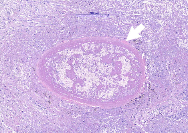
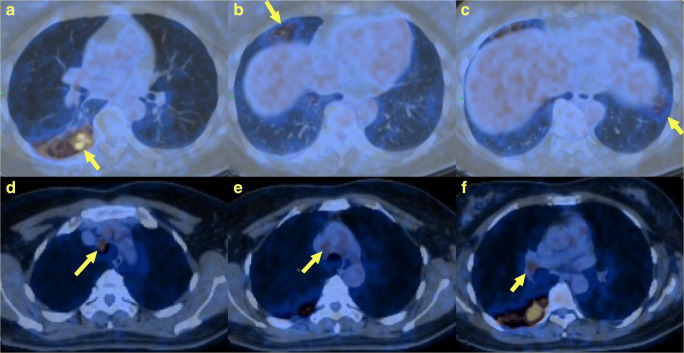
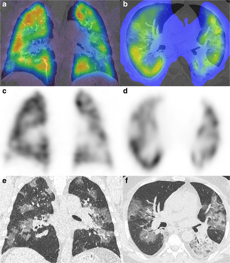
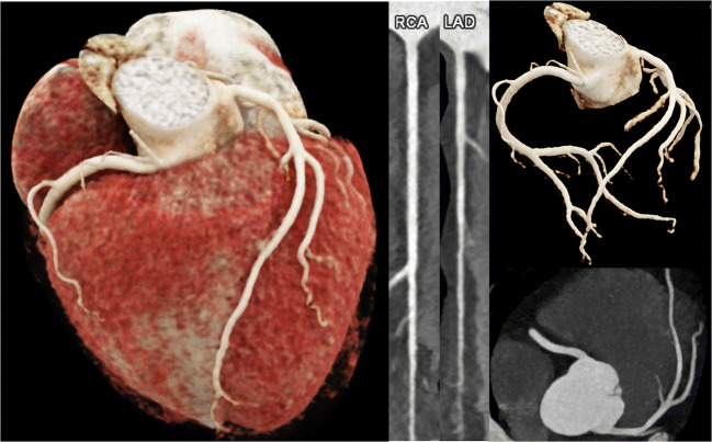
Medical imaging methods are assuming a greater role in the workup of patients with COVID-19, mainly in relation to the primary manifestation of pulmonary disease and the tissue distribution of the angiotensin-converting-enzyme 2 (ACE 2) receptor. However, the field is so new that no consensus view has emerged guiding clinical decisions to employ imaging procedures such as radiography, computer tomography (CT), positron emission tomography (PET), and magnetic resonance imaging, and in what measure the risk of exposure of staff to possible infection could be justified by the knowledge gained. The insensitivity of current RT-PCR methods for positive diagnosis is part of the rationale for resorting to imaging procedures. While CT is more sensitive than genetic testing in hospitalized patients, positive findings of ground glass opacities depend on the disease stage. There is sparse reporting on PET/CT with [18F]-FDG in COVID-19, but available results are congruent with the earlier literature on viral pneumonias. There is a high incidence of cerebral findings in COVID-19, and likewise evidence of gastrointestinal involvement. Artificial intelligence, notably machine learning is emerging as an effective method for diagnostic image analysis, with performance in the discriminative diagnosis of diagnosis of COVID-19 pneumonia comparable to that of human practitioners.

Keywords: COVID-19; Corona virus; Imaging; SARS-CoV-2.


Conflict of interest statement

The authors declare no competing interests.

Figures

Fig. 1
Fig. 1
Electron transmission micrograph showing an alveolar type II cell with its characteristic lamellar bodies (arrowheads) and a group of coronaviruses (circle)
Fig. 2
Fig. 2
Micrograph of a hematoxylin and eosine-stained lung tissue sample of a patient with severe COVID-19 pneumonia showing a small sized artery filled with a fibrin-rich thrombus with signs of organization
Fig. 3
Fig. 3
Three different patients with COVID-19 related abnormalities in the lungs shown by non-contrast CT. a A 55-year-old male patient with COVID-19 diagnosed 1 day before. CT showing ill-defined ground glass nodules in both lungs (yellow arrows). b A 49-year-old male patient with acute dyspnea for 4 days. CT showing extensive ill-defined consolidations with positive air-bronchograms in both lower lobes: COVID-19 was confirmed by RT-PCR. c A 60-year-old male patient with consolidations in the lung periphery sparing the subpleural space. The findings are compatible with organizing pneumonia indicating ongoing repair of the known COVID-19 pneumonia
Fig. 4
Fig. 4
Axial CT section at the level of the lower lobes in a patient 4 months after severe COVID-19 pneumonia. Focal, subpleural reticulations with associated volume loss (image a, arrow) and subpleural bands (arrowheads in a) represent residual fibrous foci. Axial minimal intensity projections (10-mm slice thickness; image b) picture peripheral traction bronchiectasis. In addition, a common finding in post-COVID-19 patients is multifocal air-trapping (asterisk in b) pointing towards involvement of small airways
Fig. 5
Fig. 5
A 48-year-old female was hospitalized with fever, cough, and fatigue with a high index of clinical suspicion for COVID-19. [18F]-FDG PET/CT revealed high levels of uptake in the lower lobe of the right lung (a), the middle lobe (b), and in the left lower lobe (c) with patchy opacities. High uptake was also observed in mediastinal lymph-node stations 2R (d), 4R (e), and in the right hilum (f), as shown by the yellow arrows
Fig. 6
Fig. 6
Perfusion SPECT/CT images of a heterogenous perfusion pattern in a case of COVID-19 pneumonia. Shown are the hybrid images (a/b), the perfusion SPECT (c/d), and the non-contrast enhanced CT images (e/f)
Fig. 7
Fig. 7
The heart of a 64-year-old female patient who had survived a SARS-CoV-2 infection 4 months previously. Now suffering from dyspnea and chest pain. cvRF, dyslipidemia. Results: CaSc 0, no stenosis
Fig. 8
Fig. 8
A 49-year-old male patient. New onset of dyspnea NYHA III since a SARS-CoV-2 infection 5 months ago. Cardiac MRI at 3T showing a dilated, eccentrically hypertrophied left ventricle (LV-EDV 151 ml/m2, LV mass 127 g/m2). Left ventricular (LV) ejection fraction (LV-EF 37%), LV peak circumferential (−12%), radial (18%), and longitudinal strain (−14%) were globally reduced, while the lowest regional peak circumferential strain was found in the LV septum (−6%, asterisk, a). Mid-ventricular Late-Gadolinium-Enhancement (LGE, b, c) was present in the LV septum, with normal T2 relaxation time (40 ms, d) in T2 mapping, but prolonged T1 relaxation time (1330 ms, e, f), consistent with myocardial fibrosis (white arrows)
Fig. 9
Fig. 9
Coronal T2-WI MR image in a patient after COVID-19 and hyposmia demonstrates bilateral atrophy of the olfactory bulb (yellow circle)
Fig. 10
Fig. 10
a, b A 44-year-old female patient with COVID-19 and severe ARDS. Axial FLAIR MR images show bilateral, ill-defined hyperintensity of the white matter consistent with leukoencephalopathy (white arrows)

References

    1. Xu X, et al. Imaging and clinical features of patients with 2019 novel coronavirus SARS-CoV-2. Eur J Nucl Med Mol Imaging. 2020;47(5):1275–1280. - PMC - PubMed
    1. Tang L, et al. COVID-19: a review of what radiologists need to know. World J Clin Cases. 2020;8(22):5501–5512. - PMC - PubMed
    1. Minamimoto R, et al. FDG-PET/CT images of COVID-19: a comprehensive review. Glob Health Med. 2020;2(4):221–226. - PMC - PubMed
    1. Fields BKK, et al. Imaging of COVID-19: CT, MRI, and PET. Semin Nucl Med. 2020;S0001-2998(20):30123–30129. - PMC - PubMed
    1. Galea N, et al., How to perform a cardio-thoracic magnetic resonance imaging in COVID-19: comprehensive assessment of heart, pulmonary arteries, and lung parenchyma. Eur Heart J Cardiovasc Imaging, 2020. - PMC - PubMed