. 2021 Sep;164(1):135-147.
doi: 10.1111/imm.13349. Epub 2021 May 24.

Development of a high-sensitivity ELISA detecting IgG, IgA and IgM antibodies to the SARS-CoV-2 spike glycoprotein in serum and saliva

Affiliations

Development of a high-sensitivity ELISA detecting IgG, IgA and IgM antibodies to the SARS-CoV-2 spike glycoprotein in serum and saliva

Sian E Faustini et al. Immunology. 2021 Sep.

Abstract

Detecting antibody responses during and after SARS-CoV-2 infection is essential in determining the seroepidemiology of the virus and the potential role of antibody in disease. Scalable, sensitive and specific serological assays are essential to this process. The detection of antibody in hospitalized patients with severe disease has proven relatively straightforward; detecting responses in subjects with mild disease and asymptomatic infections has proven less reliable. We hypothesized that the suboptimal sensitivity of antibody assays and the compartmentalization of the antibody response may contribute to this effect. We systematically developed an ELISA, optimizing different antigens and amplification steps, in serum and saliva from non-hospitalized SARS-CoV-2-infected subjects. Using trimeric spike glycoprotein, rather than nucleocapsid, enabled detection of responses in individuals with low antibody responses. IgG1 and IgG3 predominate to both antigens, but more anti-spike IgG1 than IgG3 was detectable. All antigens were effective for detecting responses in hospitalized patients. Anti-spike IgG, IgA and IgM antibody responses were readily detectable in saliva from a minority of RT-PCR confirmed, non-hospitalized symptomatic individuals, and these were mostly subjects who had the highest levels of anti-spike serum antibodies. Therefore, detecting antibody responses in both saliva and serum can contribute to determining virus exposure and understanding immune responses after SARS-CoV-2 infection.

Keywords: COVID-19; ELISA; SARS-CoV-2; antibodies.


Conflict of interest statement

AC and SH are employed by the Binding Site Group Ltd. AH has a commercial relationship with Binding Site Group Ltd. MTD and MG have a commercial relationship with Abingdon Health. The rest of the authors declared no conflict of interest.

Figures
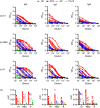
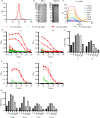
FIGURE 1
FIGURE 1
Hospitalized patients respond strongly to multiple viral proteins. Serological responses from hospitalized (HS, n = 6), non‐hospitalized convalescents (NHC, n = 5), RT‐PCR+asymptomatic subjects (AS, n = 6) or pre‐2019 normal donors (Pre19, n = 6) as determined by ELISA using HRP‐labelled anti‐IgG, IgA and IgM, against 0·1 µg purified (a) viral spike protein S1 fragment (S1), (b) receptor binding domain (RBD) or (c) nucleocapsid (N). (d) Area under the curve [11] of responses shown in a–c. The mean ± standard deviation of the mean (SD) is plotted
FIGURE 2
FIGURE 2
Stabilized, trimeric S antigen is a superior antigen to detect Ab in NHC. (a) Size‐exclusion chromatogram (SEC) for SARS‐CoV‐2 S protein fractions collected for further use and denoted by dashed grey lines. (b) Coomassie‐stained SDS‐PAGE gel for two separate expressions of SARS‐CoV‐2 (left) and silver stain of batch 1 under reducing and non‐reducing conditions (right). (c) Surface plasmon resonance (SPR) characterizing the interaction between SARS‐CoV‐2 S protein and Ace2. The plotted lines represent the averages of three analytical repeats at each concentration. (d) Serological responses from hospitalized (HS, n = 5) or pre‐2019 normal donors (Pre19, n = 6) as determined by ELISA using HRP‐labelled anti‐IgG represented as absorbance values or (e) signal:noise ratio at each serum dilution against 0·1 µg purified viral trimeric spike protein (S) or the S1 fragment (S1). (f) Mean absorbance values of 4 sera per group against 0·1 or 0·2 µg S or nucleocapsid (N). (g) Signal:noise ratio at each serum dilution against 0·1 or 0·2 µg of S or N. Error bars represent standard deviation from the mean (SD)
FIGURE 3
FIGURE 3
Antigen targeting and antibody isotypes do not differ depending upon the severity of disease. Serological responses from hospitalized (H, n = 3), non‐hospitalized convalescent (NHC, n = 3) or pre‐2019 donors (Pre19, n = 2) as determined by ELISA using HRP‐labelled anti‐IgG1, IgG2, IgG3 or IgG4 against 0·1 µg (a) trimeric spike protein (S), (b) receptor binding domain (RBD) or (c) nucleocapsid (N). (d) Area under the curve [11] of IgG1 and IgG3 responses as shown in a–c. The mean ± standard deviation of the mean (SD) is plotted
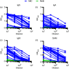
FIGURE 4
FIGURE 4
Combined detection of IgG, IgA and IgM enhances discrimination of infected and Pre19 groups. Serological responses from non‐hospitalized convalescents (NHC, n = 20) or pre‐2019 donors (Pre19, n = 4) as determined by ELISA using HRP‐labelled (a) anti‐IgG, (b) IgA and (c) IgM or (d) combined GAM, against 0·1 µg purified viral spike protein (S)
FIGURE 5
FIGURE 5
Serum and salivary anti‐SARS‐CoV‐2 antibody responses exhibit a moderate correlation. Absorbance values of paired serum diluted 1:40 and saliva diluted 1:2 from RT‐PCR‐positive convalescent healthcare workers (n = 80) who were symptomatic at the time of testing serum, as determined by ELISA using combined HRP‐labelled anti‐IgG, IgA and IgM [23] for serum and unlabelled combined anti‐IgG, IgA and IgM with a tertiary HRP‐labelled goat anti‐mouse Ig amplification step for saliva against 0·1 µg trimeric spike protein (S) or 0·2 µg nucleocapsid (N). (a) Correlation between paired serum and saliva absorbance values with percentages of samples positive for anti‐S antibodies in either serum, saliva or both. Positivity was determined by cut‐offs for each fluid (dotted lines) based on the mean +2 standard deviations of 8 pre‐2019 (Pre19) negative samples for sera and 83 pre‐2019 negative samples for saliva. Solid line represents simple linear regression of all samples. (b) Correlation between paired serum and saliva absorbance values with percentages of samples positive for anti‐N antibodies in either serum, saliva or both. Positivity was determined by cut‐offs for each fluid (dotted lines) based on the mean +2 standard deviations of 8 pre‐2019 (Pre19) negative samples for sera and 83 pre‐2019 negative samples for saliva. Solid line represents simple linear regression of all samples. (c) Correlation between paired saliva absorbance values with percentages of samples positive for anti‐S and anti‐N antibodies in saliva. Positivity was determined by cut‐offs for saliva (dotted lines) based on the mean +2 standard deviations of 83 pre‐2019 negative samples for saliva. Solid line represents simple linear regression of all samples. (d) Correlation between paired saliva absorbance values with percentages of samples positive for anti‐S and anti‐N antibodies in serum. Positivity was determined by cut‐offs for serum (dotted lines) based on the mean +2 standard deviations of 8 pre‐2019 negative samples for serum. Solid line represents simple linear regression of all samples

Update of

References

    1. WHO . Coronavirus disease (COVID‐19) Situation Report ‐ 141. 2020.
    1. Park SW, Cornforth DM, Dushoff J, Weitz JS. The time scale of asymptomatic transmission affects estimates of epidemic potential in the COVID‐19 outbreak. Epidemics. 2020;31:100392. - PMC - PubMed
    1. Shields A, Faustini SE, Perez‐Toledo M, Jossi S, Aldera E, Allen JD, et al. SARS‐CoV‐2 seroprevalence and asymptomatic viral carriage in healthcare workers: a cross‐sectional study. Thorax. 2020;75(12):1089–1094. - PMC - PubMed
    1. Woo PC, Lau SK, Wong BH, Chan KH, Hui WT, Kwan GS, et al. False‐positive results in a recombinant severe acute respiratory syndrome‐associated coronavirus (SARS‐CoV) nucleocapsid enzyme‐linked immunosorbent assay due to HCoV‐OC43 and HCoV‐229E rectified by Western blotting with recombinant SARS‐CoV spike polypeptide. J Clin Microbiol. 2004;42:5885–8. - PMC - PubMed
    1. Wibroe PP, Helvig SY, Moein MS. The role of complement in antibody therapy for infectious diseases. Microbiol Spectr. 2014;2:1–9. - PubMed

Publication types