. 2021 May 13;184(10):2680-2695.e26.
doi: 10.1016/j.cell.2021.04.001. Epub 2021 Apr 30.

LanCLs add glutathione to dehydroamino acids generated at phosphorylated sites in the proteome

Affiliations

LanCLs add glutathione to dehydroamino acids generated at phosphorylated sites in the proteome

Kuan-Yu Lai et al. Cell. .

Abstract

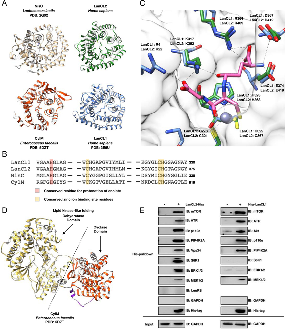
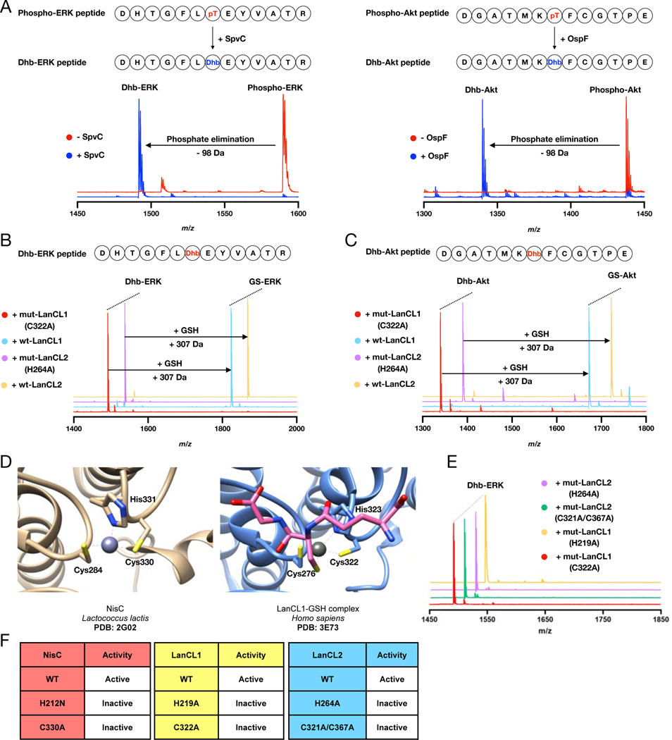
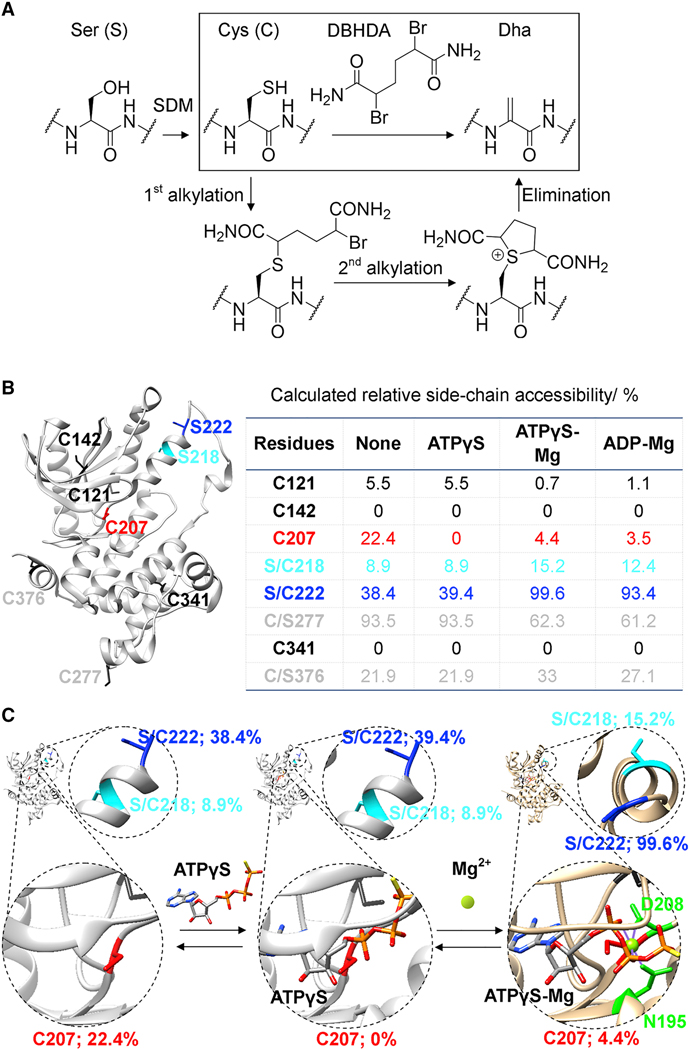
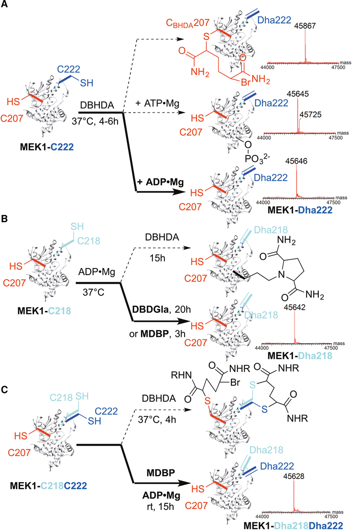
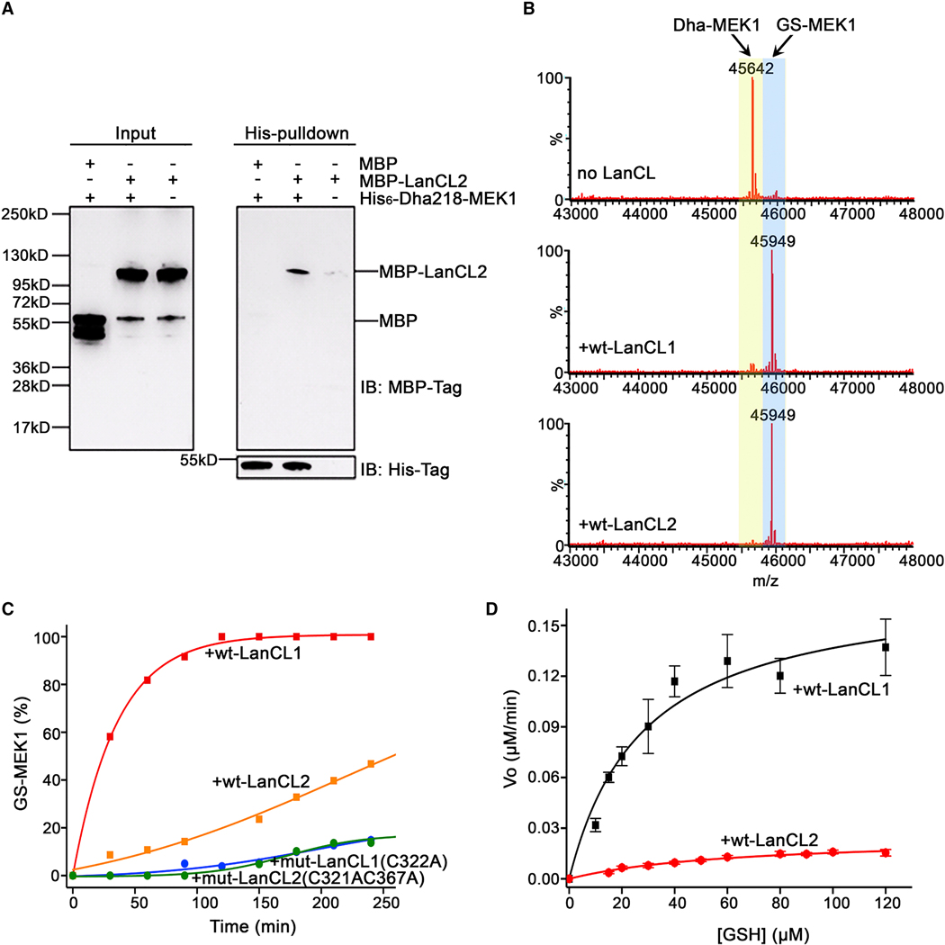
Enzyme-mediated damage repair or mitigation, while common for nucleic acids, is rare for proteins. Examples of protein damage are elimination of phosphorylated Ser/Thr to dehydroalanine/dehydrobutyrine (Dha/Dhb) in pathogenesis and aging. Bacterial LanC enzymes use Dha/Dhb to form carbon-sulfur linkages in antimicrobial peptides, but the functions of eukaryotic LanC-like (LanCL) counterparts are unknown. We show that LanCLs catalyze the addition of glutathione to Dha/Dhb in proteins, driving irreversible C-glutathionylation. Chemo-enzymatic methods were developed to site-selectively incorporate Dha/Dhb at phospho-regulated sites in kinases. In human MAPK-MEK1, such "elimination damage" generated aberrantly activated kinases, which were deactivated by LanCL-mediated C-glutathionylation. Surveys of endogenous proteins bearing damage from elimination (the eliminylome) also suggest it is a source of electrophilic reactivity. LanCLs thus remove these reactive electrophiles and their potentially dysregulatory effects from the proteome. As knockout of LanCL in mice can result in premature death, repair of this kind of protein damage appears important physiologically.

Keywords: C-glutathionylation; LanCL; MEK1; dehydroalanine; dehydrobutyrine; eliminylome; lanthionine; phosphoThr lyase; protein damage.


Conflict of interest statement

Declaration of interests The authors declare no competing interests.

Figures

Figure 1.
Figure 1.. Structural and pulldown analyses of LanC-type domains and LanCL proteins reveal common architectures and affinity for kinases.
(A) Crystal structures of human LanCLs, bacterial NisC (PDB ID 2G0D), and a LanC-domain in a LanM (CylM; PDB ID 5DZT). (B) Partial sequence alignment of NisC, the cyclase domain of CylM, and human LanCLs. The residues proposed to protonate the enolate intermediate during Michael-type addition to dehydroamino acids are in red. Zinc ion binding residues are in yellow. (C) GSH binding pocket is conserved in LanCL1 and LanCL2 but not LanC/LanMs. The apo structure of Δ1–18-LanCL2 (green sticks) was superimposed with the LanCL1-GSH co-crystal structure (blue sticks; PDB: 3E73). Arg22 is not observed in the electron density of LANCL2. Pink sticks show GSH. (D) Crystal structure of CylM showing interaction of a LanC-type cyclase domain (red) with a kinase-like dehydratase domain (gold). (E) His-pulldown assay with LanCL proteins. Recombinant His-tagged human LanCL1 and LanCL2 show affinity for several cellular kinases from HEK293 cell extracts. For pulldown of endogenous LanCL2 with a subset of these kinases, see Figure S1.
Figure 2.
Figure 2.. Eliminative damage in peptides catalyzed by the pathogen pThr lyases SpvC or OspF and addition of GSH to these eliminated peptides by wt LanCL1 and LanCL2 but not mutants.
(A) MALDI-TOF mass spectra of phospho-ERK peptide treated with His6-SpvC and phospho-Akt peptide treated with His6-OspF resulting in a loss of 98 Da. See Supplemental Table 2 for calculated and observed masses and Figure S1C for fragmentation data. (B) MALDI-TOF mass spectra of Dhb-containing ERK peptide treated with wt LanCL1- and 2 or mutants in the presence of GSH. (C) MALDI-TOF mass spectra of Dhb-containing Akt peptide treated with wt LanCL1 and LanCL2 or mutants. (D) Active sites of LanCL1 bound to GSH (PDB ID 3E73) and the intra-molecular C-S bond forming catalyst NisC (PDB ID 2G0D). (E,F) Loss of catalytic activity upon mutation reveals the importance of the His proposed to protonate the enolate during intramolecular nisin cyclase CS-bond-forming activity as well as intermolecular LanCL-catalyzed C-glutathionylation. The zinc ion binding Cys residues are also essential for LanCL activity.
Figure 3.
Figure 3.. A strategy for chemical generation of eliminated MEK1 proteins.
(A) Overall sequence for site-selective incorporation of Dha at regulatory Ser sites in kinases. Reaction of the most reactive Cys over less reactive Cys was used to allow chemical, regioselective incorporation of Dha at different sites. SDM, site directed mutagenesis. (B) MEK1, its six native free Cys (black, grey, red) and two activating Ser sites 218 (cyan) and 222 (blue); Table: Predicted side-chain accessibility of sites with/without ‘masking’ nucleotide and Mg(II); (C) Schematic of predicted structural and accessibility effects of ‘masking’ and enhancing ligands; Cys207 is blocked by ATPγS; 218 & 222 are more accessible with Mg(II). See also Figure S2.
Figure 4.
Figure 4.. Reagent-controlled, regioselective, chemical elimination reactions of MEK1 allow single site and double site elimination.
Switching of reagents and conditions selectively avoids unwanted, competing chemical pathways (dotted) and yields clean chemical elimination (bold) to desired proteins: (A) MEK1-Dha222 (B) MEK1-Dha218, or (C) MEK1-Dha218Dha222. Intact protein LC-ESI-MS shown; see Supplemental Table 2 for calculated and observed masses, Figure S2 for structures of reagents, and Figures S3–S5 for all data.
Figure 5.
Figure 5.. LanCL proteins catalyze non-canonical C-glutathionylation of dehydroamino acid containing proteins.
(A) In vitro binding analysis of MBP-LanCL2 with His6-MEK1-Dha218 by His-pulldown assay. (B) LanCL1 and LanCL2 (2 μM) C-glutathionylate His6-MEK1-Dha218. The conversions of Dha-MEK1 (2 μM) to GS-MEK1 after 40-min incubation with 1.0 mM GSH at 25 °C (Dha-MEK1: LanCL = 1:1) are shown as deconvoluted LC mass spectra. (C) Time course of GSH (2.5 mM) addition to MEK1-Dha218 (6 μM) catalyzed by LanCL1/2 (1.2 μM). The formation of GS-MEK1 was monitored according to the intensities of each product from deconvoluted spectra on LCMS. (D) Pseudo-single substrate Michaelis-Menten plot in the presence of LanCL1 or LanCL2 (Dha-MEK1, 6 μM; LanCL1/2, 1.2 μM). All kinetic assays were performed in triplicate. Initial reaction velocity at different GSH concentrations was calculated by linear regression fit to the Michaelis-Menten equation of the formation of GS-MEK1 over time using OriginPro9.7 for apparent kinetic parameters KM and kcat. For spectra and data with Dhb-Erk2 see Fig. S7; for kinase activity of Dha- and GS-MEK1, see Fig. S6.
Figure 6.
Figure 6.. LanCL-catalyzed C-glutathionylation shows flexible substrate selectivity, high stereoselectivity, is the major C-glutathionylation activity in cells, and under certain circumstances the absence of LanCL proteins leads to lethality.
(A, B, C) LanCL2, but not an active site variant, adds GSH to (A) ProcA 2.8 mut, (B) CylLS”, and (C) CylLL”. Eliminated (Dha or Dhb) residues are highlighted in red; Cys residues involved in thioether linkages are in green. (D) Comparative activity of LanCL2-His6 and His6-GSTA4 with Dhb-containing ERK peptide. (E) Non-canonical glutathionylation was observed with His6-MEK1-Dha222 when incubated with wt MEF cell extracts. No glutathionylation was observed when His6-MEK1-Dha222 was treated with cell extract of LanCL1–3 triple knockout (TKO) MEF cells. (F) Structure of DL- and LL-MeLan. (G) GC-MS analysis of the methyllanthionine in glutathionylated Dhb-ERK peptide. Deep blue: DL-MeLan synthetic standard; Red: LL-MeLan synthetic standard; Cyan: derivatized residue from GS-ERK peptide; Co-injection of derivatized MeLan from the GS-ERK peptide with DL standard (orange) or LL standard (pink). (H) Kaplan-Meier survival curve of WT (n = 58) and LanCL TKO (n = 47) FVB mice. Censored individual mice are shown as a tick mark (see STAR methods). Gehan-Breslow-Wilcoxon test showed a P value of 0.0004. Statistical significance was accepted at P < 0.05.

References

    1. Alessi DR, Saito Y, Campbell DG, Cohen P, Sithanandam G, Rapp U, Ashworth A, Marshall CJ, and Cowley S. (1994). Identification of the sites in MAP kinase kinase-1 phosphorylated by p74raf-1. EMBO J 13, 1610–1619. - PMC - PubMed
    1. Arbibe L, Kim DW, Batsche E, Pedron T, Mateescu B, Muchardt C, Parsot C, and Sansonetti PJ (2007). An injected bacterial effector targets chromatin access for transcription factor NF-kappaB to alter transcription of host genes involved in immune responses. Nat Immunol 8, 47–56. - PubMed
    1. Bauer H, Mayer H, Marchler-Bauer A, Salzer U, and Prohaska R. (2000). Characterization of p40/GPR69A as a peripheral membrane protein related to the lantibiotic synthetase component C. Biochem Biophys Res Commun 275, 69–74. - PubMed
    1. Bose AK, and Janes KA (2013). A high-throughput assay for phosphoprotein-specific phosphatase activity in cellular extracts. Mol Cell Proteomics 12, 797–806. - PMC - PubMed
    1. Brennan DF, and Barford D. (2009). Eliminylation: a post-translational modification catalyzed by phosphothreonine lyases. Trends Biochem Sci 34, 108–114. - PubMed

Publication types

MeSH terms