Review
. 2021 Jul:43:101975.
doi: 10.1016/j.redox.2021.101975. Epub 2021 Apr 20.

Glutaredoxin: Discovery, redox defense and much more

Affiliations
Review

Glutaredoxin: Discovery, redox defense and much more

Fernando T Ogata et al. Redox Biol. 2021 Jul.

Abstract

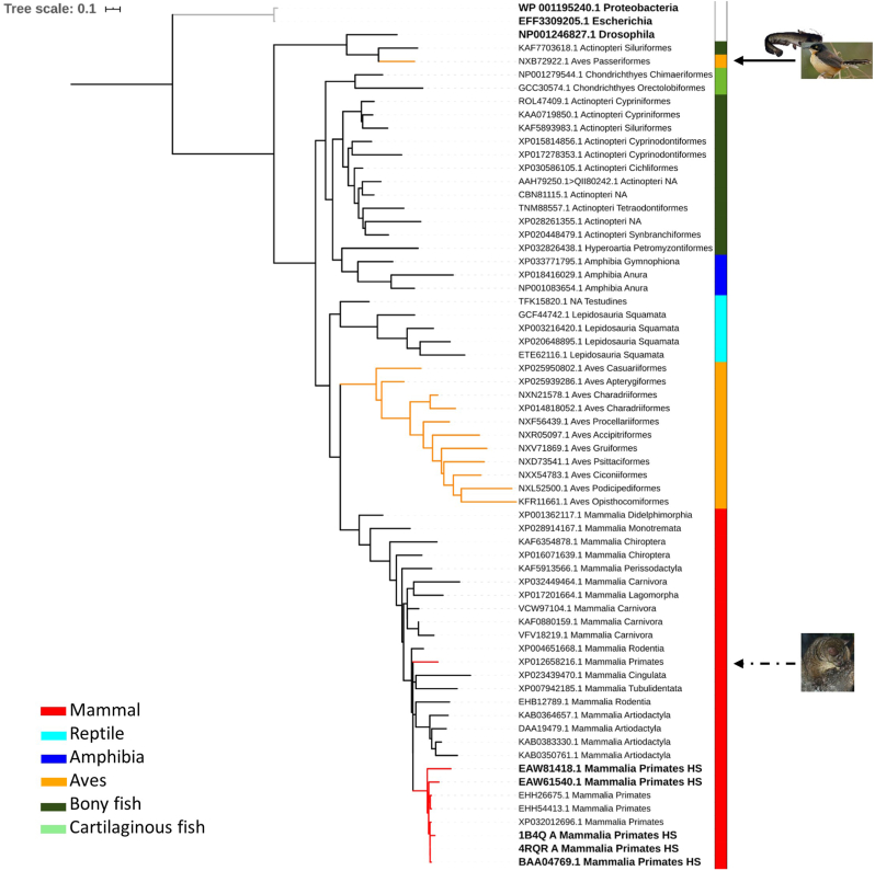
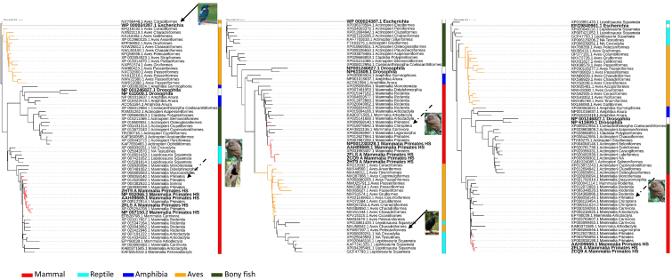
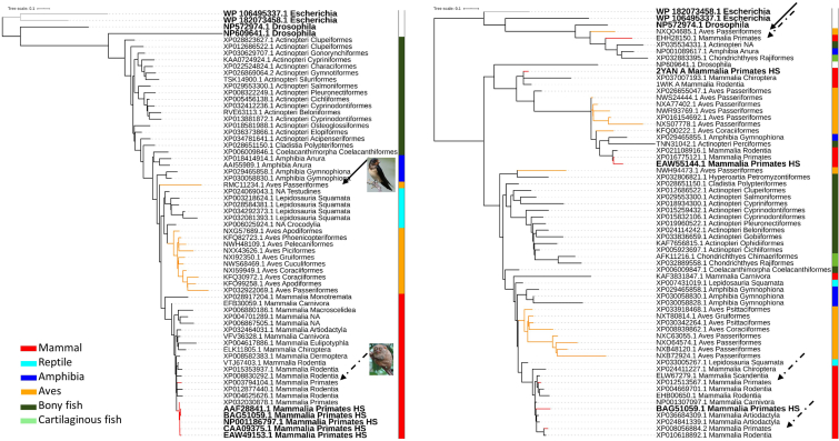
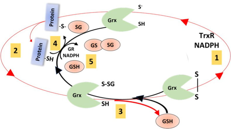
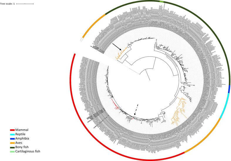
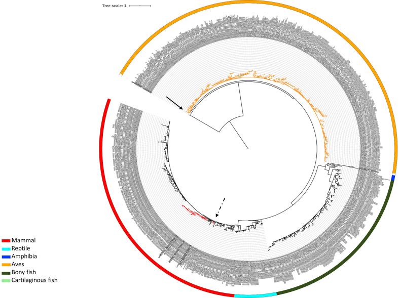
Glutaredoxin, Grx, is a small protein containing an active site cysteine pair and was discovered in 1976 by Arne Holmgren. The Grx system, comprised of Grx, glutathione, glutathione reductase, and NADPH, was first described as an electron donor for Ribonucleotide Reductase but, from the first discovery in E.coli, the Grx family has impressively grown, particularly in the last two decades. Several isoforms have been described in different organisms (from bacteria to humans) and with different functions. The unique characteristic of Grxs is their ability to catalyse glutathione-dependent redox regulation via glutathionylation, the conjugation of glutathione to a substrate, and its reverse reaction, deglutathionylation. Grxs have also recently been enrolled in iron sulphur cluster formation. These functions have been implied in various physiological and pathological conditions, from immune defense to neurodegeneration and cancer development thus making Grx a possible drug target. This review aims to give an overview on Grxs, starting by a phylogenetic analysis of vertebrate Grxs, followed by an analysis of the mechanisms of action, the specific characteristics of the different human isoforms and a discussion on aspects related to human physiology and diseases.

Keywords: Deglutathionylation; Glutaredoxin; Glutathionylation; Grxs phylogenetics; Iron homeostasis; Redox regulation.


Conflict of interest statement

The authors report no conflict of interest.

Figures

Fig. 1
Fig. 1
Maximum-likelihood phylogenetic tree of Grx1 protein sequences of representative species of vertebrates classes. Bold taxa indicate human or outgroup glutaredoxins. Red tree nodes indicate primates, and yellow nodes indicate bird’s glutaredoxins. Grey nodes indicate the E. coli outgroup. Vertebrate classes are colored coded by the lateral strip. Continuous black arrows indicate heterogeneous bird clades, and discontinuous back arrows indicate heterogeneous primate’s clades, with images of the species in question. A collection of sequences was retrieved from Genbank after performing a BLASTP [228] of Grx1 (NP_001112362.1 glutaredoxin-1 [Homo sapiens]). The number of sequences retrieved after BLASTP search varied between 500 and 1000, using as criterion having at least one species belonging to Mammal, Bird (Aves), Bony/Cartilaginous fish, Reptile and Amphibia. Each taxon in the phylogenetic tree is displayed by the accession number and the taxonomic class and order. Human sequences are identified with HS. Additionally, since Grxs are ubiquitous enzymes [17], we included E. coli and Drosophila melanogaster Grxs as outgroups, choosing the two most similar sequences found by BLASTP for each of the Grxs. Each protein sequence was aligned using MAFFT version 7 [229]. Identical sequences were removed before constructing maximum-likelihood phylogenetic trees with FastTreeMP [230]. The Interactive Tree of Life (iTOL) v4 [231] was used to visualize and annotate trees. To reduce the tree complexity, the leaves that contribute the least to the tree diversity were pruned using Treemmer v0.2 [232]. (For interpretation of the references to color in this figure legend, the reader is referred to the Web version of this article.)
Fig. 2
Fig. 2
Maximum-likelihood phylogenetic tree of Grx2 protein sequences of representative species of vertebrates classes. Bold taxa indicate human or outgroup glutaredoxins. Isoforms 1, 2 and 3 from the left to the right. Red tree nodes indicate primates, and yellow nodes indicate bird’s glutaredoxins. Grey nodes indicate the E. coli outgroup. Vertebrate classes are colored coded by the lateral strip. Continuous black arrows indicate heterogeneous bird clades, and discontinuous back arrows indicate heterogeneous primate’s clades, with images of the species in question. A collection of sequences was retrieved from Genbank after performing a BLASTP [228] of Grx2 isoform 1 (NP_057150.2 glutaredoxin 2 isoform 1 [Homo sapiens]; Grx2b), Grx2 isoform 2 (NP_932066.1 glutaredoxin 2 isoform 2 precursor [Homo sapiens]; Grx2a), Grx2 isoform 3 (NP_001230328.1 glutaredoxin 2 isoform 3 [Homo sapiens]; Grx2c) and the phylogenetic tree constructed in a similar manner as for Fig. 2. (For interpretation of the references to color in this figure legend, the reader is referred to the Web version of this article.)
Fig. 3
Fig. 3
Maximum-likelihood phylogenetic tree of Grx3 protein sequences of representative species of vertebrates classes. Bold taxa indicate human or outgroup glutaredoxins. Isoforms 1 and 2 from the left to the right. Red tree nodes indicate primates, and yellow nodes indicate bird’s glutaredoxins. Grey nodes indicate the E. coli outgroup. Vertebrate classes are colored coded by the lateral strip. Continuous black arrows indicate heterogeneous bird clades, and discontinuous back arrows indicate heterogeneous primate’s clades, with images of the species in question. A collection of sequences was retrieved from Genbank after performing a BLASTP [228] of Grx3 isoform 1 (NP_006532.2 glutaredoxin-3 isoform 1 [Homo sapiens]), Grx3 isoform 2 (NP_001308909.1 glutaredoxin-3 isoform 2 [Homo sapiens]) and the phylogenetic tree constructed in a similar manner as for Fig. 2. (For interpretation of the references to color in this figure legend, the reader is referred to the Web version of this article.)
Fig. 4
Fig. 4
Maximum-likelihood phylogenetic tree of Grx5 protein sequences of representative species of vertebrates classes. Bold taxa indicate human or outgroup glutaredoxins. Red tree nodes indicate primates, and yellow nodes indicate bird’s glutaredoxins. Grey nodes indicate the E. coli outgroup. Vertebrate classes are colored coded by the lateral strip. Continuous black arrows indicate heterogeneous bird clades, with images of the species in question A collection of sequences was retrieved from Genbank after performing a BLASTP [228] of Grx5 (NP_057501.2 glutaredoxin-related protein 5, mitochondrial precursor [Homo sapiens]) and the phylogenetic tree constructed in a similar manner as for Fig. 2. (For interpretation of the references to color in this figure legend, the reader is referred to the Web version of this article.)
Fig. 5
Fig. 5
Sequence Logos of the active sites of glutaredoxins present in vertebrates obtain from a multiple sequence alignment of 500 to 1000 glutaredoxins sequences. Active site described for each glutaredoxin. Consensus: most common amino acid found in the alignment. Sequence Logo representing the conservation at each position. Identity: Green: 100% identity, Greenish-brown: at least 30% and under 100% identity, Red: below 30% identity. Sequences logos were produced from multiple sequence alignments, where the height of the amino acid symbol represents its relative frequency and the height of the stack represents the sequence conservation at each position [233]. B) Representation of the tridimensional surface and secondary structure of human glutaredoxins evidencing the active site in yellow (Protein Data Base structures 1B4Q, 2CQ9, 2DIY and 2MMZ for GRX1, GRX2, GRX3 and GRX5, respectively) visualized using PyMOL 2.4. (For interpretation of the references to color in this figure legend, the reader is referred to the Web version of this article.)
Fig. 6
Fig. 6
Schematic representation of Grx catalytic mechanisms. (A) Monothiol mechanism of Grx. This mechanism depends only on the N-terminal active site cysteinyl residue that attacks the GSH moiety (1) and forms a GSH-mixed disulfide intermediate (2). Thus, the substrate is reduced. A molecule of GSH is required to reduce the Grx-S-SG mixed disulfide (3) Electrons are transferred from NADPH via GR to reduce GSSG (4) (B) Dithiol mechanism of Grx. This mechanism depends on both active site cysteines. The N-terminal active site cysteine has a low pKa value, allowing the initiation of a nucleophilic attack preferentially on mixed disulfides with GSH with formation of a covalently bound mixed disulfide intermediate (1). In the second step the C-terminal active site cysteine reduces the mixed disulfide releasing the reduced protein (2). The oxidized Grx is reduced by two molecule of GSH (3 and 4) electrons are transferred from NADPH via GR to reduce GSSG (5) Grx: glutaredoxin; GSH: reduced glutathione; GSSG: oxidized glutathione; GR: glutathione reductase; NADPH: nicotinamide adenine dinucleotide phosphate.
Fig. 7
Fig. 7
Schematic representation of Grx2 activity. This scheme shows how Grx2 catalyses the glutathionylation and deglutathionylation of protein thiols. The red circle represents alternative way of Grx2 reduction, via TrxR and NADPH (1) and PSSG reduction forming glutathionylated Grx2 (2). The second cysteine in the active site remove the GSH form the Grx2 leading to oxidized protein (3). However, glutathionylated Grx2 can transfer the GSH to a target protein (4 black arrow). Grx2 canonical dithiol mechanism is represented by step 2 (red arrow) and 5 (black arrow), mechanism explained in more details in Fig. 6B (Figure based on [28]). (For interpretation of the references to color in this figure legend, the reader is referred to the Web version of this article.)
figs1
figs1
figs2
figs2
figs3
figs3
figs4
figs4
figs5
figs5

References

    1. Holmgren A. Hydrogen donor system for Escherichia coli ribonucleoside diphosphate reductase dependent upon glutathione. Proc. Natl. Acad. Sci. U.S.A. 1976;73:2275–2279. doi: 10.1073/pnas.73.7.2275. - DOI - PMC - PubMed
    1. Laurent T.C., Moore E.C., Reichard P. Enzymatic Synthesis of Deoxyribonucleotides. Iv. Isolation and and characterization of thioredoxin, the hydrogen donor from Escherichia coli b. J. Biol. Chem. 1964;239:3436–3444. http://www.ncbi.nlm.nih.gov/pubmed/14245400 - PubMed
    1. Fernandes A.P., Holmgren A. Glutaredoxins: glutathione-dependent redox enzymes with functions far beyond a simple thioredoxin backup system. Antioxidants Redox Signal. 2004;6:63–74. doi: 10.1089/152308604771978354. - DOI - PubMed
    1. Vilella F., Alves R., Rodríguez-Manzaneque M.T., Bellí G., Swaminathan S., Sunnerhagen P., Herrero E. Evolution and cellular function of monothiol glutaredoxins: involvement in iron-sulphur cluster assembly. Comp. Funct. Genom. 2004;5:328–341. doi: 10.1002/cfg.406. - DOI - PMC - PubMed
    1. Mondal S., Kumar V., Singh S.P. Phylogenetic distribution and structural analyses of cyanobacterial glutaredoxins (Grxs), Comput. Biol. Chem. 2020;84 doi: 10.1016/j.compbiolchem.2019.107141. - DOI - PubMed

Publication types

LinkOut - more resources