

Ocular manifestations of ectodermal dysplasia
- PMID: 33933124
- PMCID: PMC8088613
- DOI: 10.1186/s13023-021-01824-2
Ocular manifestations of ectodermal dysplasia
Abstract
Purpose: The ectodermal dysplasias (EDs) constitute a group of disorders characterized by abnormalities in two or more ectodermal derivatives, including skin, hair, teeth, and sweat glands. The purpose of the current study was to evaluate ocular manifestations in pediatric patients with ED.
Methods: Retrospective case series including consecutive ED subjects who were treated in the ophthalmology department at the Children's Hospital of Philadelphia over a 12-year period (2009-2020). Main Outcome Measures were ocular and ocular adnexal abnormalities.
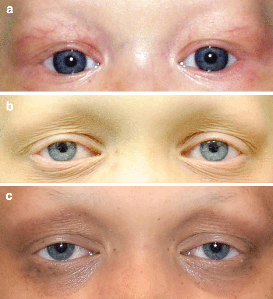
Results: Thirty subjects were included: 20 males (67%), mean age of 4.5 years (range 0.3-18). Patients with different subtypes were included, with the hypohidrotic ED and ectrodactyly-ectodermal dysplasia-clefting variants being most prevalent. Most common findings were: lacrimal drainage obstruction in 12 (40%) including punctal agenesis in 10 (33%), refractive errors in 13 (43%) and amblyopia in 6 (20%). A new finding of eyelid ptosis or eyelash ptosis was demonstrated in 11 subjects (37%), mostly associated with TP63 or EDA1 genes variants.
Conclusion: Ectodermal dysplasias are associated with various ocular pathologies and amblyopia in the pediatric population. We report a possible genetic association between lash ptosis and EDA1 gene, and eyelid ptosis and TP63 or EDA1 genes variants.
Keywords: AEC; Ankyloblepharon-ectodermal defects-cleft lip/palate; EDA1; EEC; Ectodermal dysplasia; Ectrodactyly-ectodermal dysplasia-clefting; Lash ptosis; Ptosis; TP63.
Conflict of interest statement
None.
Figures
References
-
- Jen M, Nallasamy S. Ocular manifestations of genetic skin disorders. Clin Dermatol. 2016;34:242–275. - PubMed
-
- Ansari A, Pillarisetty LS. Embryology, Ectoderm. In: StatPearls. Treasure Island (FL): StatPearls Publishing; 2020. Available at: http://www.ncbi.nlm.nih.gov/books/NBK539836/ [Accessed June 1, 2020]. - PubMed
-
- Priolo M. Ectodermal dysplasias: an overview and update of clinical and molecular-functional mechanisms. Am J Med Genet A. 2009;149A:2003–2013. - PubMed
-
- Itin PH, Fistarol SK. Ectodermal dysplasias. Am J Med Genet C Semin Med Genet. 2004;131C:45–51. - PubMed
MeSH terms
LinkOut - more resources
Full Text Sources
Other Literature Sources
Medical
