. 2022 Dec;164(6):e411-e424.
doi: 10.1016/j.jtcvs.2021.03.100. Epub 2021 Apr 9.

Allergic response to medical products in patients with alpha-gal syndrome

Affiliations

Allergic response to medical products in patients with alpha-gal syndrome

Kasinath V Kuravi et al. J Thorac Cardiovasc Surg. 2022 Dec.

Abstract

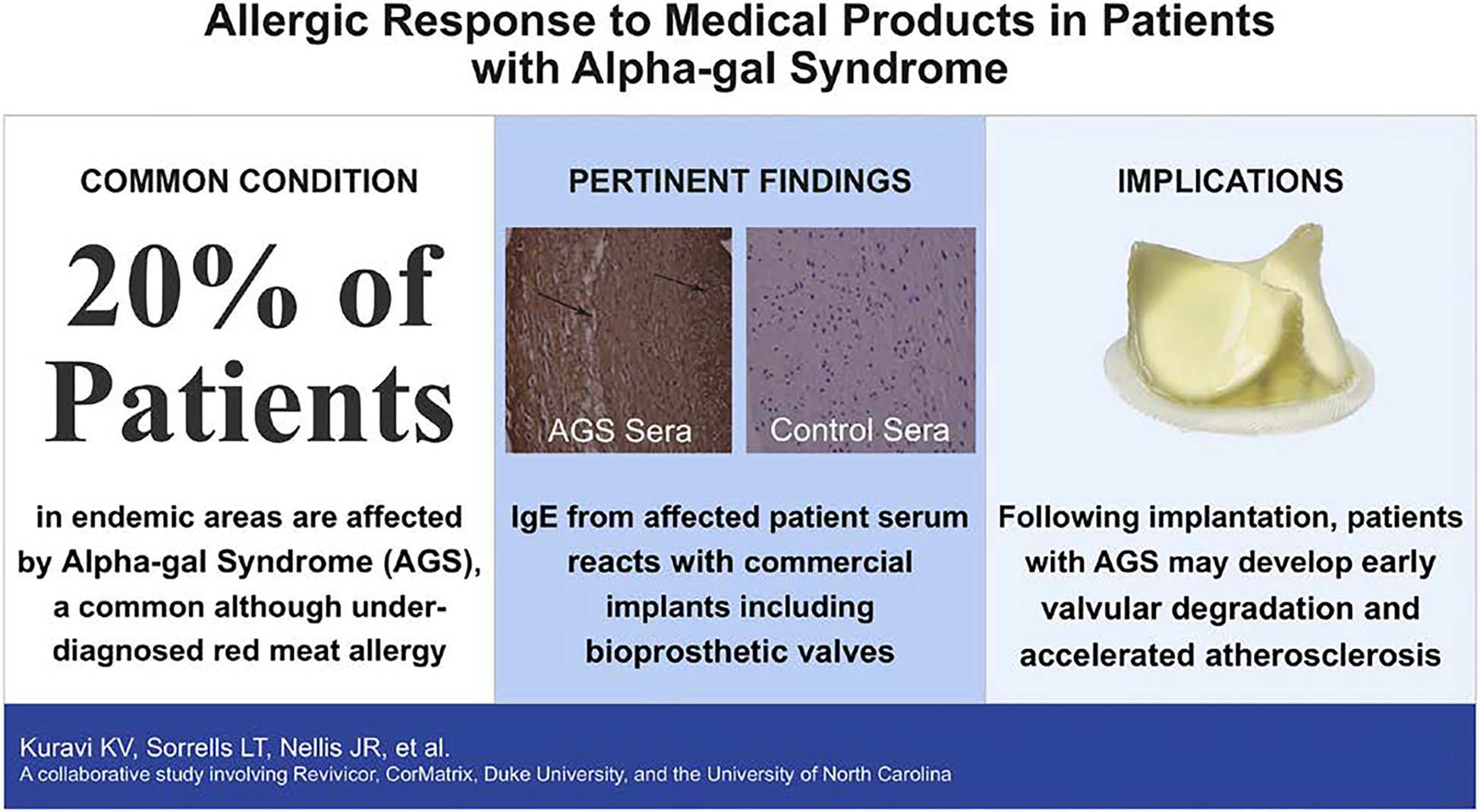
Background: Galactose-α-1,3-galactose (alpha-gal) is a carbohydrate that is ubiquitously expressed in all mammals except for primates and humans. Patients can become sensitized to this antigen and develop alpha-gal syndrome (AGS), or a red meat allergy. Symptoms range from generalized gastroenteritis and malaise to anaphylaxis, and in endemic areas, the prevalence can be as high as 20%. Although AGS patients commonly avoid alpha-gal by avoiding meat, patients have also developed symptoms due to animal-derived medical products and devices. With the rise in transcatheter aortic valve replacement, we investigate the immunogenicity of common cardiac materials and valves.

Objective: To assess the in vitro immunoglobulin E response toward common medical products, including cardiac patch materials and bioprosthetic valves in patients with AGS.

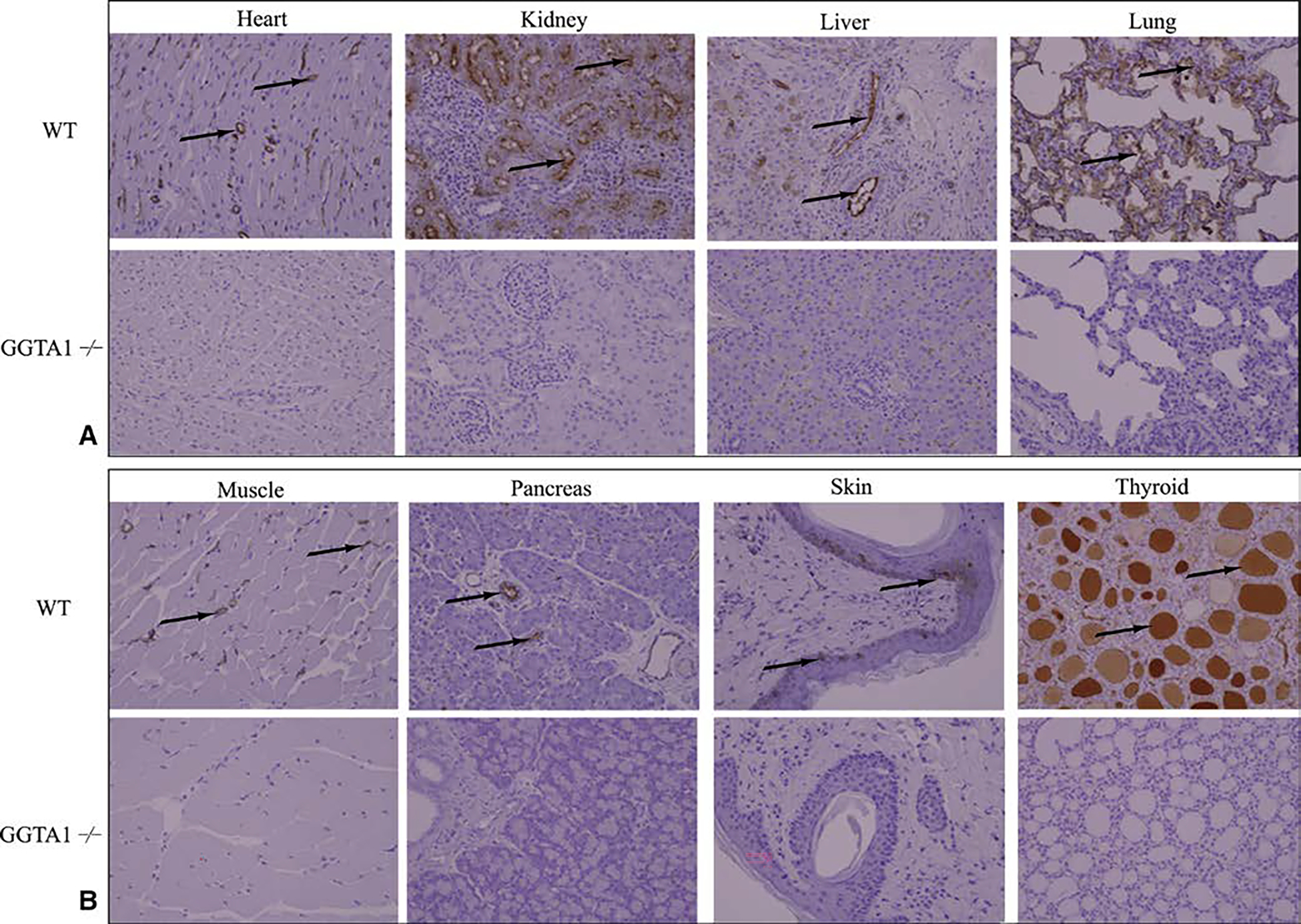
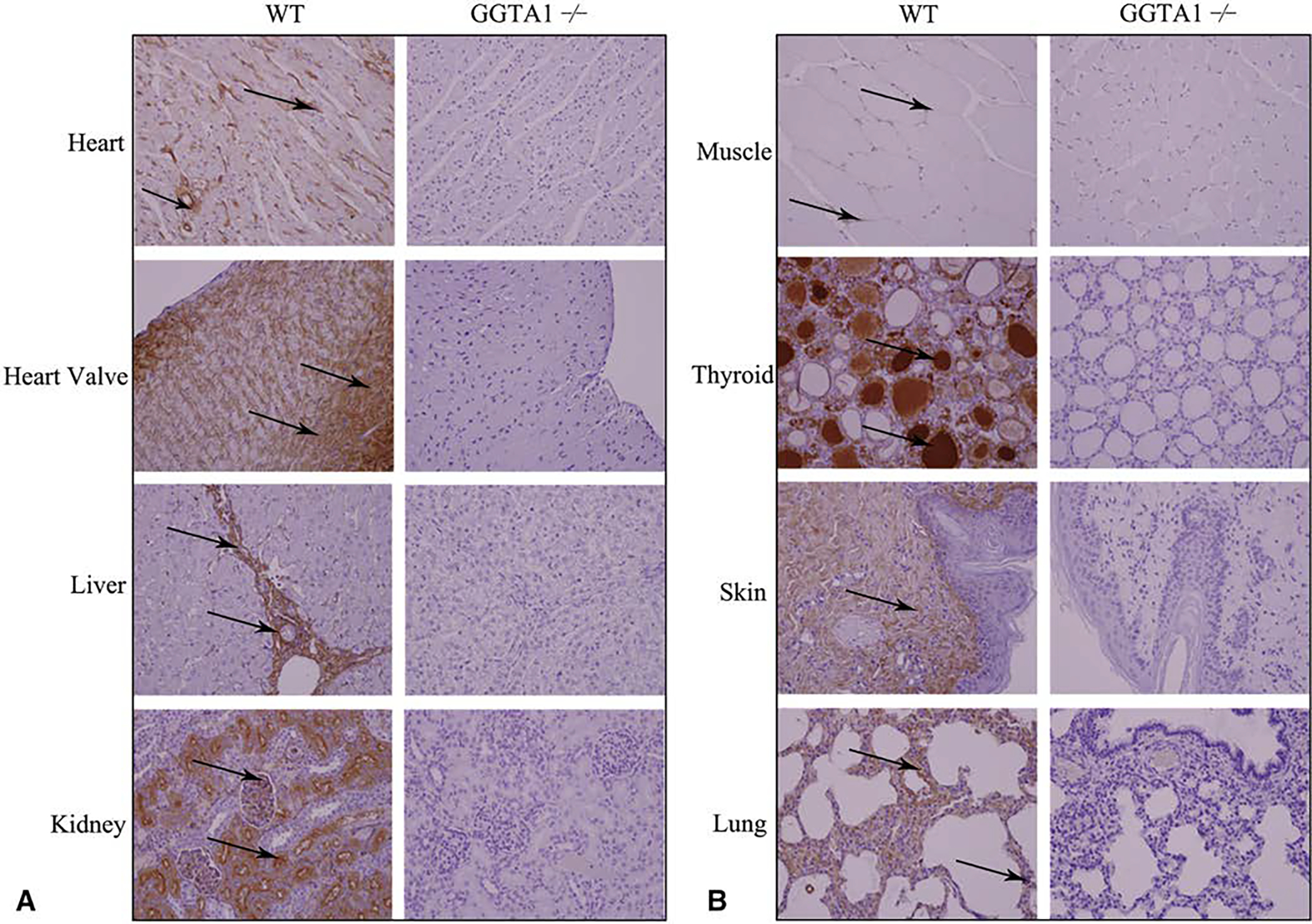
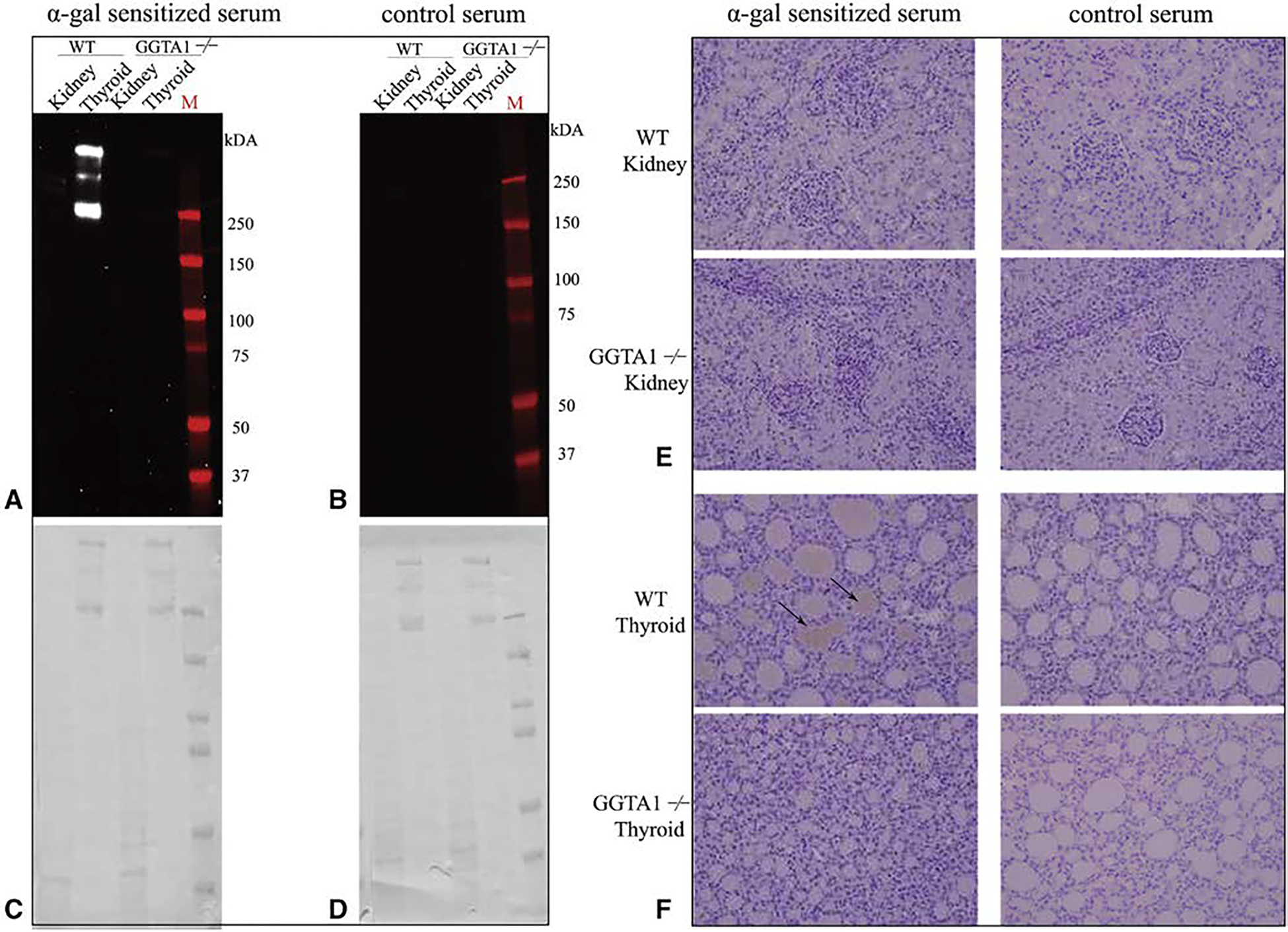
Methods: Immunoblot and immunohistochemistry techniques were applied to assess immunoglobulin E reactivity to various mammalian derived tissues and medical products for patients with AGS.

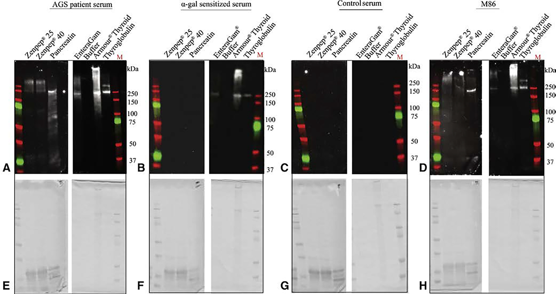
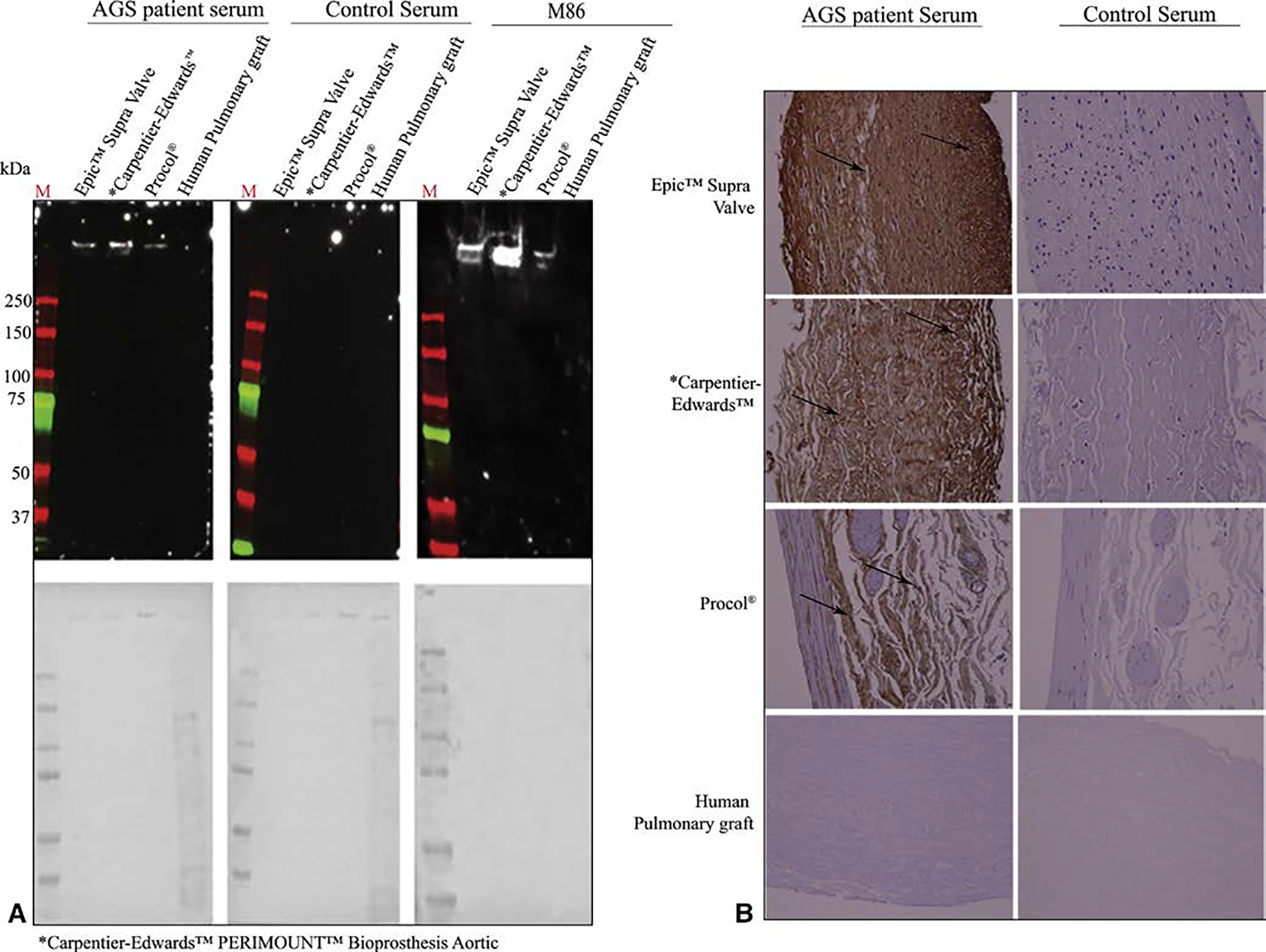
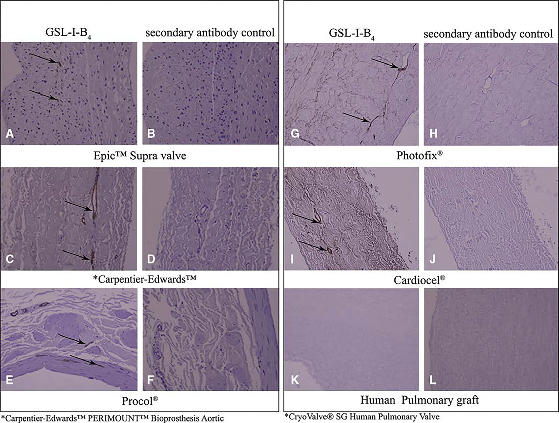
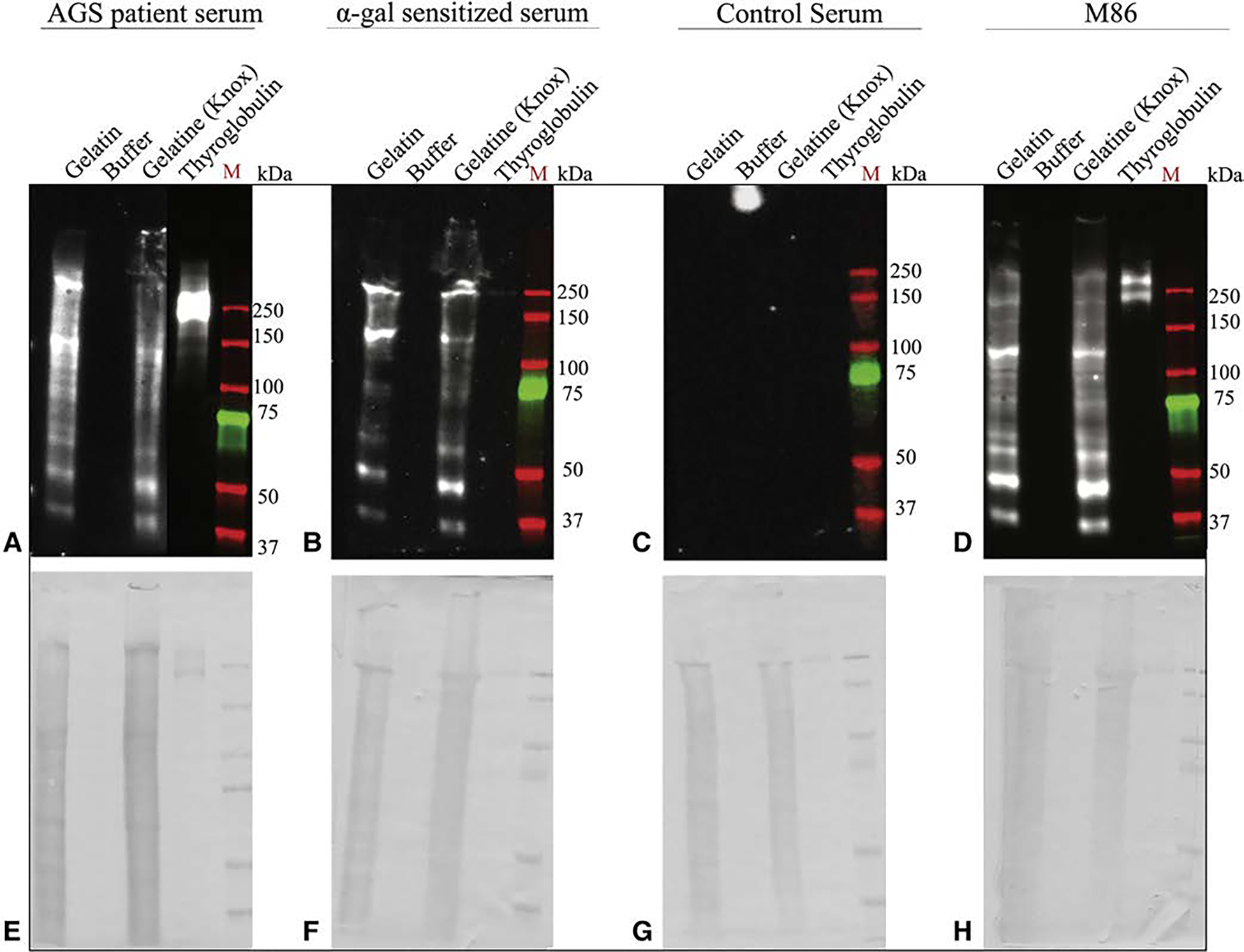
Results: AGS serum showed strong reactivity to all of the commercially available, nonhuman products tested, including various decellularized cardiac patch materials and bioprosthetic aortic valves. AGS serum did not react to tissues prepared using alpha-gal knockout pigs.

Conclusions: Despite commercial decellularization processes, alpha-gal continues to be present in animal-derived medical products, including bioprosthetic valves. Serum from patients with AGS demonstrates a strong affinity for these products in vitro. This may have serious potential implications for sensitized patients undergoing cardiac surgery, including early valve failure and accelerated coronary artery disease.

Keywords: allergy; alpha-gal; alpha-gal syndrome; bioprosthetic valve; chronic inflammation; coronary artery disease; early valve degradation.


Conflict of interest statement

Conflict of Interest Statement

The authors reported no conflicts of interest.

The Journal policy requires editors and reviewers to disclose conflicts of interest and to decline handling or reviewing manuscripts for which they may have a conflict of interest. The editors and reviewers of this article have no conflicts of interest.

Figures

FIGURE 1.
FIGURE 1.
Alpha-gal immunoglobulin E (IgE) reactivity toward commercially available drugs. Immunoblot analysis for alpha-gal IgE reactivity toward Zenpep (Allergan, Irvine, Calif), EnteraGam (Entera Health, Cary, NC), Armour Thyroid (Allergan), and thyroglobulin. A, Using alpha-gal syndrome (AGS) patient serum. B, Using alpha-gal sensitized serum. C, Using control serum from volunteers without AGS. D, Using M86, an alpha-gal antibody. E through H, The associated Coomassie stains indicating the presence of protein.
FIGURE 2.
FIGURE 2.
Alpha-gal immunoglobulin E (IgE) reactivity toward commercially available cardiac patch materials. Materials used include Photofix (Cryolife, Kennesaw, Ga), Cardiocel (LeMaitre Vascular, Burlington, Mass), CorMatrix Cor Patch 1.0, and CorMatrix Cor Patch 2.0 (CorMatrix Cardiovascular Inc, Sunnyvale, Calif). A, Immunoblots using alpha-gal syndrome (AGS) patient serum, M86 (an alpha-gal antibody), and control serum from patients without AGS. Assays using serum were counterstained for human IgE B, Immunohistochemisty of the given products using serum from patients with and without AGS. All images are at 200×. Brown staining is indicative of alpha-gal IgE.
FIGURE 3.
FIGURE 3.
Alpha-gal immunoglobulin E (IgE) reactivity toward commercially available bioprosthetic aortic valves and vascular grafts. Materials used include the Epic Supra Valve (Abbott, Abbott Park, Ill), Carpentier-Edwards Perimount Valve (Edwards Lifesciences, Irvine, Calif), Procol Bovine Mesenteric Vein Conduit (LeMaitre Vascular, Burlington, Mass), and CryoValve SG Human Pulmonary Valve Conduit (Cryolife, Kennesaw, Ga). A, Immunoblots using alpha-gal syndrome (AGS) patient serum, M86 (an alpha-gal antibody), and control serum from patients without AGS. Assays using serum were counterstained for human IgE B, Immunohistochemisty of the given products using serum from patients with and without AGS. All images are at 200×. Brown staining is indicative of alpha-gal IgE.
FIGURE 4.
FIGURE 4.
Immunohistochemistry for alpha-gal using commercially available surgical implants. Labels for an associated product are located below the panel. Products included in the figure include Epic Supra Valve (Abbott Park, Ill), Carpentier-Edwards Perimount Valve (Edwards Lifesciences, Irvine, Calif), Procol Bovine Mesenteric Vein Conduit (LeMaitre Vascular, Burlington, Mass), Photofix (Cryolife, Kennesaw, Ga), Cardiocel (LeMaitre Vascular), and CryoValve SG Human Pulmonary Valve Conduit (Cryolife). A, C, E, G, and I, Brown staining (black arrows) indicates the presence of alpha-gal using griffonia simplicifolia lectin I – isolectin B4 (GSL I-B4), which binds alpha-galactose residues. B, D, F, H, J, K, and L, No staining is seen with the omission of GSL I-B4 and use of secondary antibody alone, or in the CryoValve SG Human Pulmonary Valve Conduit, which serves as a negative control. All images at 200×.
FIGURE 5.
FIGURE 5.
Alpha-gal immunoglobulin E (IgE) reactivity toward commercially available gelatin. Immunoblot analysis for alpha-gal IgE reactivity using laboratory grade gelatin and commercial grade Gelatine (Knox Company, Parsipanny, NJ). A, Alpha-gal syndrome (AGS) patient serum, B, Alpha-gal sensitized serum. C, Control serum from volunteers without AGS. D, M86, an alpha-gal antibody. E though H, The associated Coomassie stains indicating the presence of protein.
FIGURE 6.
FIGURE 6.
In summary, alpha-gal syndrome (AGS) is a common red meat allergy. Sensitized patients develop an immunoglobulin E (IgE) response to alpha-gal, a carbohydrate that is expressed by all lesser mammals, including cows and pigs. This is particularly important for patients undergoing bioprosthetic valve replacement. As shown, patients with AGS react to the bioprosthetic valve (brown alpha-gal IgE staining based on patient sera), whereas healthy control sera does not (right panel). The consequences of this may include chronic inflammation, early valve degradation, and accelerated atherosclerosis.
FIGURE E1.
FIGURE E1.
Alpha-gal immunohistochemistry for wild type (WT) and glycoprotein galactosyltransferase α-1,3 (GGTA1−/−) porcine organs. A and B, WT and GGTA1−/− tissues were stained with biotinylated griffonia simplicifolia lectin I – isolectin B4 (GSL I-B4) lectin to demonstrate the presence or absence of alpha-gal. The presence of brown pigment (black arrows) indicates a positive reaction with the alpha-gal epitope in wild type tissues. The lack of brown pigment confirms absence of alpha-gal in glycoprotein galactosyltransferase α-1,3 (GGTA1−/−) pig tissues. All images are at 200×.
FIGURE E2.
FIGURE E2.
Immunoblot analysis of alpha-gal glycosylated proteins in different porcine organs. A, Protein extracts from wild type (WT) and glycoprotein galactosyltransferase α-1,3 (GGTA1−/−) pig kidney samples were detected using alpha-gal epitope binding antibody (ie, M86). B and C, Alpha-gal binding profiles of WT, GGTA1+/−, and GGTA1−/− porcine heart, lung, kidney, heart valve, pancreas, and thyroid tissue samples using alpha-gal syndrome (AGS) patient serum. D, M86 detection of WT and GGTA1−/− liver, skin and muscle extracts. E though H, The Coomassie stains for the associated immunoblots, indicating the presence of protein. Positive signal in WT and GGTA1+/− samples indicates presence of alpha-gal glycosylation. No signal in GGTA1−/− samples indicates absence of alpha-gal glycosylation.
FIGURE E3.
FIGURE E3.
Alpha-gal immunoglobulin E (IgE) reactivity toward glycosylated proteins in different wild type (WT) and glycoprotein galactosyltransferase α-1,3 (GGTA1−/−) porcine organs. A and B, Alpha-gal binding profiles of WT and GGTA1−/− porcine heart, heart valve, liver, kidney, muscle, thyroid, skin, and lung using alpha-gal syndrome (AGS) patient sera. Positive signal (brown) represents the presence of alpha-gal IgE binding. All images are at 200×.
FIGURE E4.
FIGURE E4.
Alpha-gal immunoglobulin E (IgE) reactivity toward glycosylated proteins in different wild type (WT) and glycoprotein galactosyltransferase α-1,3 (GGTA1−/−) porcine organs. A through C, Alpha-gal binding profiles of WT and GGTA1−/− porcine heart, lung, kidney, thyroid, skin, muscle, liver, heart, heart valve, and pancreas using alpha-gal syndrome (AGS) patient serum. Positive signal represents the presence of alpha-gal glycosylated proteins. D, Alpha-gal binding profiles of WT and GGTA1−/− heart, lung, liver, pancreas, and kidney using control serum from patients without AGS. No signal is seen representing the absence of alpha-gal glycosylated proteins. E though H, The Coomassie stains for the associated immunoblots, indicating the presence of protein.
FIGURE E5.
FIGURE E5.
Alpha-gal immunoglobulin E (IgE) reactivity toward different wild type (WT) and glycoprotein galactosyltransferase α-1,3 (GGTA−/−) porcine organs in a sensitized patient. A, Sensitized patient serum binding profiles for WT and GGTA1−/− kidney and thyroid. B, Alpha-gal binding profiles of WT and GGTA1−/− kidney and thyroid using control serum from patients without elevated alpha-gal IgE. C and D, The Coomassie stains associated with the corresponding immunoblots. E and F, Immunohistochemical binding profile of alpha-gal glycosylated proteins in WT and GGTA−/− kidney and thyroid using sensitized and control serum. Brown staining represents alpha-gal IgE binding. All images are at 200×.

Comment in

Similar articles

Cited by

References

    1. Macher BA, Galili U. The Galalpha1,3Galbeta1,4GlcNAc-R (alpha-Gal) epitope: a carbohydrate of unique evolution and clinical relevance. Biochim Biophys Acta. 2008;1780:75–88. - PMC - PubMed
    1. Galili U, Mandrell RE, Hamadeh RM, Shohet SB, Griffiss JM. Interaction between human natural anti-alpha-galactosyl immunoglobulin G and bacteria of the human flora. Infect Immun. 1988;56:1730. - PMC - PubMed
    1. Chung CH, Mirakhur B, Chan E, Le QT, Berlin J, Morse M, et al. Cetuximab-induced anaphylaxis and IgE specific for galactose-alpha-1,3-galactose. N Engl J Med. 2008;358:1109–17. - PMC - PubMed
    1. Platts-Mills TAE, Commins SP, Biedermann T, van Hage M, Levin M, Beck LA, et al. On the cause and consequences of IgE to galactose-alpha-1,3-galactose: a report from the National Institute of Allergy and Infectious Diseases Workshop on understanding IgE-mediated mammalian meat allergy. J Allergy Clin Immunol. 2020;145:1061–71. - PMC - PubMed
    1. Apostolovic D, Tran TA, Starkhammar M, Sánchez-Vidaurre S, Hamsten C, Van Hage M. The red meat allergy syndrome in Sweden. Allergo J Int. 2016;25:49–54. - PMC - PubMed

Supplementary concepts