Review
. 2022 Jun:126:71-78.
doi: 10.1016/j.semcdb.2021.04.012. Epub 2021 Apr 28.

Biochemical mechanisms of period control within the mammalian circadian clock

Affiliations
Review

Biochemical mechanisms of period control within the mammalian circadian clock

Jonathan M Philpott et al. Semin Cell Dev Biol. 2022 Jun.

Abstract

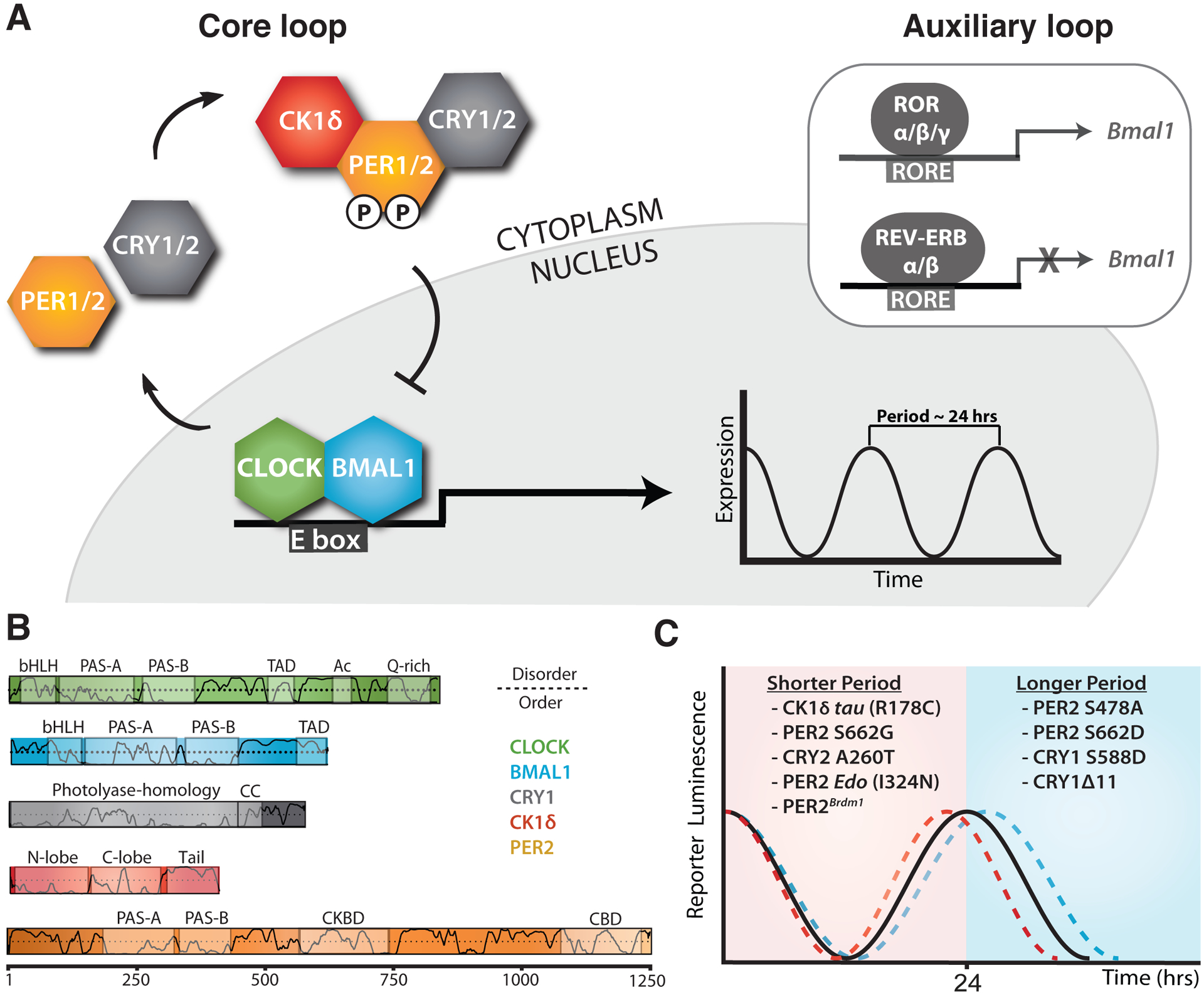
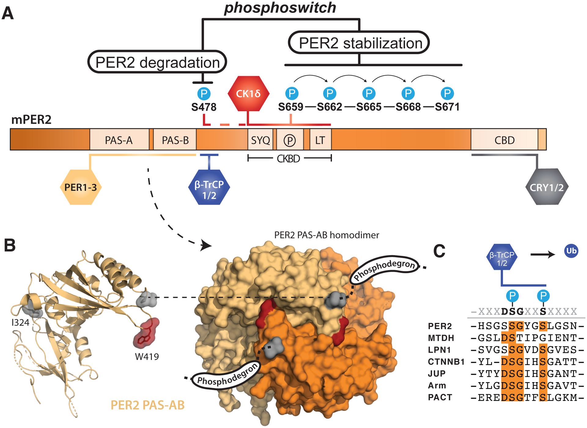
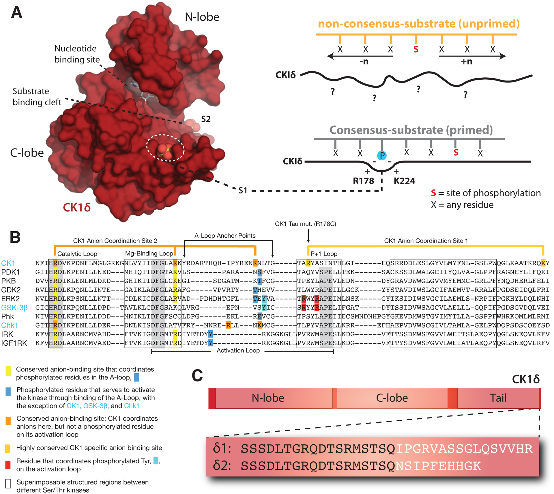
Genetically encoded biological clocks are found broadly throughout life on Earth, where they generate circadian (about a day) rhythms that synchronize physiology and behavior with the daily light/dark cycle. Although the genetic networks that give rise to circadian timing are now fairly well established, our understanding of how the proteins that constitute the molecular 'cogs' of this biological clock regulate the intrinsic timing, or period, of circadian rhythms has lagged behind. New studies probing the biochemical and structural basis of clock protein function are beginning to reveal how assemblies of dedicated clock proteins form and evolve through post-translational regulation to generate circadian rhythms. This review will highlight some recent advances providing important insight into the molecular mechanisms of period control in mammalian clocks with an emphasis on structural analyses related to CK1-dependent control of PER stability.

Keywords: Degradation; Dynamics; Feedback loop; Post-translational modifications; Proteins; Structure.


Figures

Figure 1.
Figure 1.. Changes to the core molecular clock control mammalian circadian period
(A) Simplified schematic of the core mammalian feedback loop mediated by CLOCK:BMAL1 expression of CRY and PER genes with an auxiliary loop that consists of the retinoic acid receptor-related orphan receptor α (RORα) and the nuclear receptors REV-ERBα/β. (B) Functional domain architecture of core clock proteins with structured domains (boxes) and traces indicating the propensity for intrinsic disorder [92]: bHLH, basic helix-loop-helix; PAS, PER-ARNT-SIM; TAD, transactivation domain; Ac, acetyl-CoA binding, Q-rich, polyglutamine; CC, coiled-coil; CKBD, Casein Kinase 1-binding domain; and CBD, CRY-binding domain with residue numbering underneath. (C) Period effects from select mammalian clock alleles of core clock proteins.
Figure 2.
Figure 2.. PER2 stability is tightly regulated by post-translational modifications
(A) Domain map of mouse PER2 depicting clock protein binding sites and the CK1δ-dependent phosphoswitch model. CK1δ-dependent phosphorylation of the FASP region (P) within the CKBD antagonizes activity at the upstream phosphodegron, which is used to recruit the E3 ubiquitin ligases, β-TrCP1/2, for subsequent proteasomal degradation of PER2. (B) Left, cartoon representation of the mouse PER2 PAS-AB domain monomer with the location of the Edo mutation (I324N) and Trp residue required for dimerization (W419) highlighted, PDB: 3GDI. Right, surface representation of the PER2 PAS-AB homodimer. The phosphodegron is located in a disordered region immediately downstream of the PAS-B domain; the C-terminal residue is depicted in surface mode (gray) to show how each respective phosphodegron is poised to protrude from the same face of the dimer. (C) Alignment of phosphodegrons within human proteins that are targeted by β-TrCP1/2.
Figure 3
Figure 3. Regulatory mechanisms control CK1δ activity in the mammalian clock
(A) Left, Surface representation of the CK1δ kinase domain, PDB: 5×17. The substrate binding cleft is flanked by two highly conserved anion binding sites, S1 and S2. Right, CK1 is thought to use S1 to bind phosphorylated (or ‘primed’) substrates, leading to phosphorylation of the CK1 consensus motif, pSxxS. CK1 also exhibits non-consensus activity on unprimed sites. (B) Alignment of the activation loop and nearby regions in representative Ser/Thr kinases. CK1 lacks the conserved APE motif in the P+1 loop involved in substrate recognition. (C) Differences between the alternatively spliced variants CK1δ1 and CK1δ2 at the C-terminus of the kinase.

References

    1. Aschoff J: Circadian Rhythms in Man. Science 1965, 148(3676):1427–1432. - PubMed
    1. Bass J, Lazar MA: Circadian time signatures of fitness and disease. Science 2016, 354(6315):994–999. - PubMed
    1. Takahashi JS: Transcriptional architecture of the mammalian circadian clock. Nat Rev Genet 2017, 18(3):164–179. - PMC - PubMed
    1. Aryal RP, Kwak PB, Tamayo AG, Gebert M, Chiu PL, Walz T, Weitz CJ: Macromolecular Assemblies of the Mammalian Circadian Clock. Mol Cell 2017, 67(5):770–782 e776. - PMC - PubMed
    1. Lee C, Etchegaray JP, Cagampang FR, Loudon AS, Reppert SM: Posttranslational mechanisms regulate the mammalian circadian clock. Cell 2001, 107(7):855–867. - PubMed

Publication types