. 2021 Jan-Jun:296:100730.
doi: 10.1016/j.jbc.2021.100730. Epub 2021 Apr 30.

MRCKβ links Dasm1 to actin rearrangements to promote dendrite development

Affiliations

MRCKβ links Dasm1 to actin rearrangements to promote dendrite development

Xiao-Xiao Wang et al. J Biol Chem. 2021 Jan-Jun.

Abstract

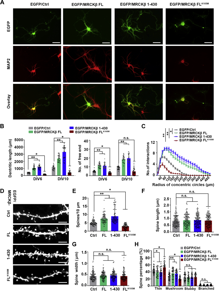
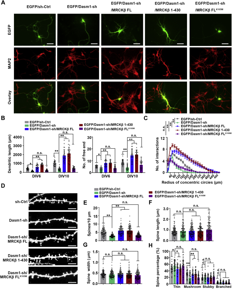
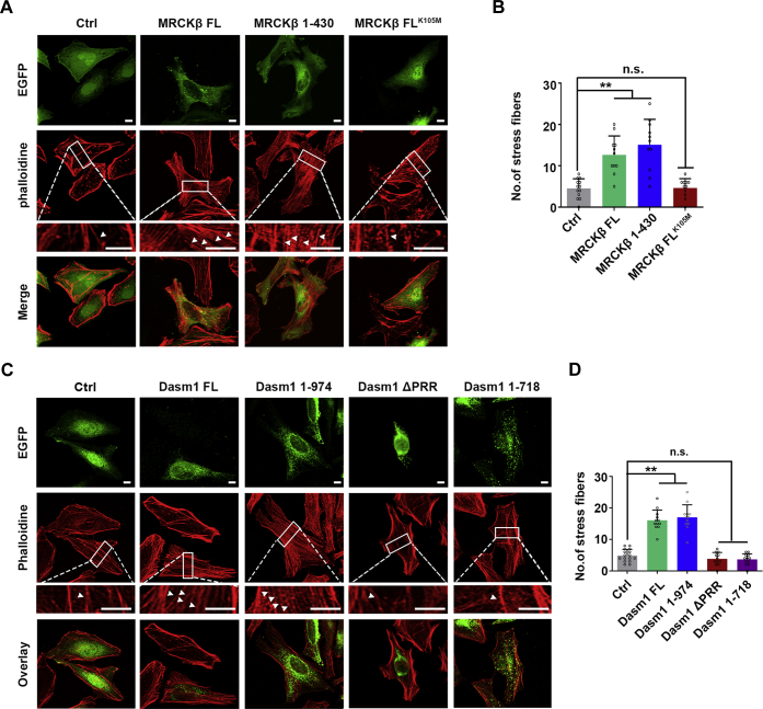
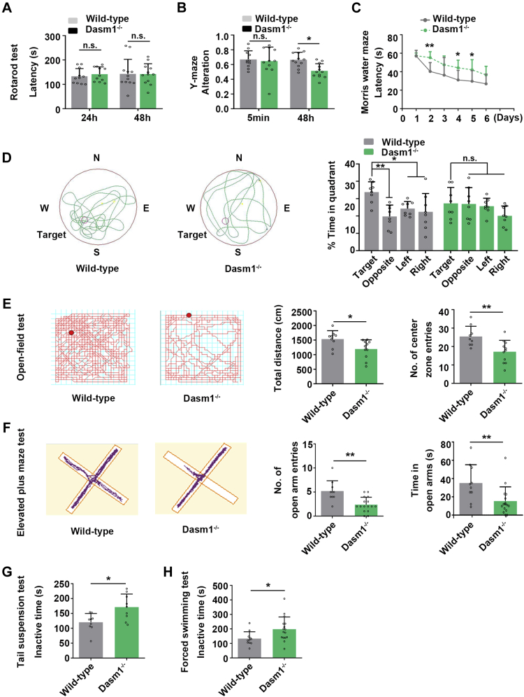
Proper dendrite morphogenesis and synapse formation are essential for neuronal development and function. Dasm1, a member of the immunoglobulin superfamily, is known to promote dendrite outgrowth and excitatory synapse maturation in vitro. However, the in vivo function of Dasm1 in neuronal development and the underlying mechanisms are not well understood. To learn more, Dasm1 knockout mice were constructed and employed to confirm that Dasm1 regulates dendrite arborization and spine formation in vivo. We performed a yeast two-hybrid screen using Dasm1, revealing MRCKβ as a putative partner; additional lines of evidence confirmed this interaction and identified cytoplasmic proline-rich region (823-947 aa) of Dasm1 and MRCKβ self-activated kinase domain (CC1, 410-744 aa) as necessary and sufficient for binding. Using co-immunoprecipitation assay, autophosphorylation assay, and BS3 cross-linking assay, we show that Dasm1 binding triggers a change in MRCKβ's conformation and subsequent dimerization, resulting in autophosphorylation and activation. Activated MRCKβ in turn phosphorylates a class 2 regulatory myosin light chain, which leads to enhanced actin rearrangement, causing the dendrite outgrowth and spine formation observed before. Removal of Dasm1 in mice leads to behavioral abnormalities. Together, these results reveal a crucial molecular pathway mediating cell surface and intracellular signaling communication to regulate actin dynamics and neuronal development in the mammalian brain.

Keywords: Dasm1; MRCKβ; actin rearrangement; dendrite development; spine formation.


Conflict of interest statement

Conflict of interest The authors declare that they have no conflicts of interest with the contents of this article.

Figures

Figure 1
Figure 1
Dasm1 knockout inhibits dendrite arborization and spine formation in mice.A, representative images of Golgi-stained hippocampal pyramidal neurons in Dasm1−/− mice and WT littermates. Scale bar, 50 μm. B, morphometric reconstruction of hippocampal pyramidal neurons in Dasm1−/− mice and WT littermates. Scale bar, 100 μm. C, Dasm1−/− mice displayed decreased total dendritic length of hippocampal pyramidal neurons fixed at postnatal (P) 5, P10, P15, and P30, but not P60, compared with WT littermates. Results were expressed as mean ± SD; n = 30 to 35; ∗p < 0.05, ∗∗p < 0.01, n.s., not significant; Unpaired two-tailed t test. D, Dasm1−/− mice displayed less branches of hippocampal pyramidal neurons fixed at P10, P15, and P30, but not P60, compared with WT littermates. Results were expressed as mean ± SD; n = 20 to 83; ∗p < 0.05, ∗∗p < 0.01, n.s., not significant; Unpaired two-tailed t test. E, Sholl analysis showed that Dasm1−/− mice displayed a reduced dendritic complexity of hippocampal pyramidal neurons at P30. Concentric circles with a 20 μm spacing were drawn around cell body, and the intersection number of all dendritic branches with the circles was counted. Results were expressed as mean ± SD; n = 20 to 83; ∗p < 0.05, ∗∗p < 0.01; two-way ANOVA (Interaction: F (18, 2983) = 70.53, p < 0.0001; Row Factor: F (18, 2983) = 8005, p < 0.0001; Column Factor: F (1, 2983) = 511.6, p < 0.0001). F, representative images of hippocampal pyramidal neurons harboring spines in Dasm1−/− mice and wild-type littermates fixed at P30. Scale bar, 5 μm. GI, quantification of spine morphology of hippocampal pyramidal neurons in Dasm1−/− mice and WT littermates. Dasm1−/− mice showed decreased spine density (G), and unchanged spine length (H), and spine width (I). Results were expressed as mean ± SD; n = 52 to 81; ∗∗p < 0.01, n.s., not significant; Unpaired two-tailed t test. J, Dasm1−/− mice showed increased thin spines and decreased mushroom spines. Results were expressed as mean ± SD; n = 30 to 36; ∗p < 0.05, n.s., not significant; two-way ANOVA (Interaction: F (3, 256) = 5.483, p = 0.0011; Row Factor: F (3, 256) = 325.7, p < 0.0001; Column factor: F (1, 256) = 6.377e-012, p > 0.9999). MRCKβ, myotonic dystrophy-related Cdc42-binding kinases beta.
Figure 2
Figure 2
Identification of MRCKβ as a Dasm1 interactor.A, confocal immunofluorescence showed co-localization of Dasm1 and MRCKβ in HeLa cells and dissociated hippocampal neurons cultured for 2 or 6 days in vitro (2 DIV or 6 DIV). Upper panel scale bar, 20 μm; Middle scale bar, 10 μm; Lower panel scale bar, 40 μm. The “% colocalization” shows the percentage of Dasm1 overlapping with MRCKβ to the total Dasm1. B, co-immunoprecipitation of endogenous Dasm1 with MRCKβ in mouse brain lysates. Mouse brain lysates were collected at postnatal (P) 7 and P 14 days and subjected to co-immunoprecipitation assay using indicated antibodies. C, co-immunoprecipitation of Dasm1 with abundant MRCKβ and little MRCKγ, but not MRCKα in 293T cells. Myc-tagged Dasm1 was co-expressed with HA-tagged MRCKα, MRCKβ, or MRCKγ in 293T cells, and cell extracts were subjected to co-immunoprecipitation assay using indicated antibodies. Results were from three independent experiments. D, Dasm1 bound to MRCKβ via its intracellular PRR. Upper panel, schematic representation of Dasm1 and various deletion mutants used for mapping Dasm1-binding site. Lower panel, HA-tagged MRCKβ was co-expressed with various Myc-tagged Dasm1 deletion mutants in 293T cells as indicated, and cell extracts were subjected to co-immunoprecipitation assay using the indicated antibodies. Text annotation in red color indicates the binding site required within Dasm1. E, MRCKβ binds to Dasm1 via its self-activating kinase domain (CC1, 410–744 amino-acid residues). Upper panel, schematic representation of MRCKβ and various truncated mutants used for mapping MRCKβ-binding site. Lower panel, Myc-tagged Dasm1 was co-expressed with various HA-tagged MRCKβ truncated mutants in 293T cells as indicated, and cell extracts were subjected to co-immunoprecipitation assay using the indicated antibodies. Text annotation in red color indicates the binding site required within MRCKβ. F, the direct interaction between Dasm1-PRR and MRCKβ-CC1 was validated by GST pull-down assay. Left panel, in vitro–translated MRCKβ was pulled down by purified GST-Dasm1-PRR fusion protein. Right panel, in vitro–translated Dasm1 was pulled down by purified GST-MRCKβ-CC1 or GST-MRCKβ-1 to 939 fusion protein. G, the three-dimensional structure of Dasm1/MRCKβ complex in stereo. The structure of MRCKβ 410 to 744 amino-acid residues was depicted in surface (green), in which the amino acid residues selected as candidate interfaces were colored with blue. The structure of the Dasm1 PRR domain was depicted in surface (magenta), in which the amino acid residues selected as candidate interfaces were colored with red. H, overview of the Dasm1/MRCKβ interface in stereo. Left panel, residues of MRCKβ (red) which polar contacted to Dasm1 (magenta) were depicted in atom-colored stick (light blue). Right panel, residues of Dasm1 (blue) which polar contacted to MRCKβ (green) were depicted in atom-colored stick (white). I, essential amino acids for the interaction between Dasm1 and MRCKβ. We generated Myc-tagged Dasm1-PRR (Myc-PRR-mut) and HA-tagged MRCKβ CC1 (HA-CC1-mut) plasmids carrying multiple point mutation based on the 3-D predictions. Myc-PRR-mut stands for Myc-Dasm1-PRR (R772A, V859A, A860G, S862A, Q863A, K865A, A911G, P932A, L934A). HA-CC1-mut stands for HA-MRCKβ CC1 (E54A, W58A, F409A, L415A, K416A, Q431A). Then, co-immunoprecipitation assay was employed to test the interaction. C1, protein kinase C conserved region 1 domain; CC, coil-coiled domain; CH, citron homology domain; CRIB, Cdc42/Rac interactive binding domain; Ig, immunoglobulin domain; FL, full-length; FNIII, Fibronectin III domain; MRCKβ, myotonic dystrophy-related Cdc42-binding kinases beta; PH, Pleckstrin homology-like domain; pPRR, preproline-rich region; PRR, proline-rich region; SP, signal peptide; TM, transmembrane domain.
Figure 3
Figure 3
Dasm1 promotes MRCKβ kinase activity.A, schematic representation of in vitro MRCKβ kinase Assay. HA-tagged MRCKβ or Myc-tagged Dasm1 was expressed in 293T cells and purified with anti-HA tagged or anti-Myc antibody-conjugated protein G coated agarose beads. The GST-MLC2 fusion protein was expressed in BL21 bacterial cells and purified using Glutathione-Sepharose 4B beads. Purified HA-MRCKβ protein alone or a combination of HA-MRCKβ and Myc-Dasm1 protein was added to the kinase reaction system, followed by MRCKβ kinase activity assay using GST-MLC2 as substrate. B, in vitro MRCKβ kinase Assay showed that HA-MRCKβ incubated with Myc-Dasm1 (lane 2, left panel; lane 2, right panel) presented higher kinase activity than HA-MRCKβ incubated alone (lane 1, left panel; lane 1, right panel). Left panel, protein used in the kinase activity assay was verified by Western blotting and Coomassie Blue Staining. Right panel, MRCKβ kinase activity was determined as the phosphorylation level of its substrate MLC2. Results were expressed as mean ± SD; n = 3; ∗p < 0.05, n.s., not significant; Unpaired two-tailed test. C, Dasm1 overexpressing promoted MLC2 phosphorylation on Ser19 and Thr18 residues in HeLa cells. D, knockout of Dasm1 inhibited MLC2 phosphorylation on Ser19 and Thr18 residues in brain lysate from mice in vivo. E, quantification of the immunoblotting results corresponding to C (left panel) and D (right panel). Results were expressed as mean ± SD; n = 3 to 4; ∗p < 0.05; ∗∗p < 0.01; Unpaired two-tailed t test. F, Dasm1 significantly inhibited intramolecular interaction between MRCKβ kinase domain (CAT) and CC autoinhibitory domain, this effect required the interaction between Dasm1 and MRCKβ CC1. Left panel, Dasm1 overexpressing inhibited the interaction between MRCKβ CAT and CC domain in HEK293T cells. Middle panel, CC1 domain is required for Dasm1-mediated inhibitory effect on intramolecular interaction between MRCKβ CAT and CC domain. Right panel, quantification of the immunoblotting results. Results were expressed as mean ± SD; n = 3; ns, not significant, ∗p < 0.05; one-way ANOVA C: (F (2, 6) = 11.00, p = 0.0098); D: (F (2, 6) = 202.7, p < 0.0001). G, Dasm1 significantly enhanced MRCKβ autophosphorylation in Dasm1-overexpressing HeLa cells. Autophosphorylation assays were carried out as described in method. Right panel, quantification of MRCKβ autophosphorylation level. Results were expressed as mean ± SD; n = 3; ∗∗p < 0.01; Unpaired two-tailed t test. H, Dasm1 significantly enhanced MRCKβ dimerization. Extracts from HeLa cells were exposed to increasing concentrations of BS3 as indicated, and cross-linked products were detected by Western blot with anti-MRCKβ antibody. Right panel, quantification of the dimer MRCKβ. Dasm1 significantly enhanced MRCKβ dimerization. Results were expressed as mean ± SD; n = 3; ∗p < 0.05, ∗∗p < 0.01, n.s., not significant; two-way ANOVA (Interaction: F (2, 12) = 5.567, p = 0.0195; Row Factor: F (2, 12) = 6.713, p = 0.0111; Column Factor: F (1, 12) = 20.51, p = 0.0007). I, the proposed “OFF-ON” switch model depicts how Dasm1 regulates MRCKβ catalytic activity. The intramolecular interaction between the Dasm1 CC autoinhibitory domain CC2-CC3 and the kinase domain keeps the kinase in a closed, inactive, and monomeric structure. The interaction between Dasm1 and MRCKβ CC1 domain opens MRCKβ closed loop and allows its N terminus–mediated dimerization, autophosphorylation, and subsequent kinase activation. MLC2, class 2 regulatory myosin light chains; MRCKβ, myotonic dystrophy-related Cdc42-binding kinases beta.
Figure 4
Figure 4
MRCKβ kinase activity is required for dendrite and spine morphogenesis.A, representative images of dendrite morphology in dissociated hippocampal neurons. Neurons were transfected with control EGFP (EGFP/Ctrl), EGFP/MRCKβ full-length (FL), EGFP/MRCKβ kinase domain (1–430), or EGFP/MRCKβ kinase-inactive K105M mutant plasmids. Neurons were cultured for 10 days in vitro and labeled with antibody against MAP2. Scale bar, 100 μm. B, quantification of total dendrite length and branches in neurons transfected with indicated plasmids. Overexpression of MRCKβ FL and MRCKβ 1 to 430 substantially increased dendrite length (left panel) and dendrite branches (right panel) at 6 DIV, 10 DIV. Overexpression of MRCKβ kinase-inactive K105M mutant significantly decreased dendrite length (left panel) and dendrite branches (right panel) at 6 DIV. Results were expressed as mean ± SD; n = 20 to 26; ∗p < 0.05, ∗∗p < 0.01, n.s., not significant; two-way ANOVA; Left panel: (Interaction: F (3, 152) = 42.09, p < 0.0001; Row Factor: F (1, 152) = 311.3, p < 0.0001; Column factor: F (3, 152) = 210.6, p < 0.0001). Right panel: (Interaction: F (3, 184) = 11.16, p < 0.0001; Row Factor: F (1, 184) = 129.7, p < 0.0001; Column factor: F (3, 184) = 155.5, p < 0.0001). C, Sholl analysis revealed that MRCKβ kinase activity is required for production of dendrite complexity at 10 DIV. Overexpression of MRCKβ FL and MRCKβ 1 to 430 substantially increased dendrite complexity, whereas overexpression of MRCKβ K105M decreased dendrite complexity. Results were expressed as mean ± SEM; n = 20 to 25; ∗p < 0.05, ∗∗p < 0.01; two-way ANOVA (Interaction: F (72, 2050) = 12.66, p < 0.0001; Row Factor: F (24, 2050) = 145.9, p < 0.0001; Column factor: F (3, 2050) = 783.8, p < 0.0001). D, representative images of dendritic spines in 25 DIV neurons transfected with indicated plasmids. Scale bar, 5 μm. E, quantification analysis of spine density in neurons transfected with indicated plasmids. Spine density was increased in neurons transfected with MRCKβ FL and MRCKβ 1 to 430, whereas decreased in neurons transfected with MRCKB K105M. Results were expressed as mean ± SD; n = 80; ∗∗p < 0.01; Kruskal-Wallis test (F (3, 316) = 87.47, p < 0.0001). F and G, spine length (F) and width (G) were not altered in neurons transfected with full-length MRCKβ or various MRCKβ mutant plasmids. Results were expressed as mean ± SD; n = 114 to 125; n.s., not significant; Kruskal-Wallis test; (F): F (3, 496) = 0.5189, p = 0.6694). (G): F (3, 452) = 0.8247, p = 0.4807. H, overexpression of MRCKβ FL and MRCKβ 1 to 430 significantly decreased the portion of thin spines and increased the portion of mushroom spines, whereas overexpression of MRCKB K105M increased the portion of thin spines and decreased the portion of mushroom spines. Results were expressed as mean ± SD; n = 30 to 35; ∗p < 0.05, ∗∗p < 0.01, n.s., not significant; two-way ANOVA (Interaction: F (9, 480) = 11.56, p < 0.0001; Row Factor: F (3, 480) = 418.3, p < 0.0001; Column factor: F (3, 480) = 1.325e-029, p > 0.9999). DIV, day in vitro; MRCKβ, myotonic dystrophy-related Cdc42-binding kinases beta.
Figure 5
Figure 5
MRCKβ rescues dendrite growth defects caused by Dasm1 suppression.A, representative images of dendrite morphology in dissociated hippocampal neurons. Neurons were transfected with control EGFP (EGFP/sh-Ctrl), EGFP/Dasm1-shRNA (sh), and various rescue plasmids including EGFP/MRCKβ full length (FL), EGFP/MRCKβ kinase domain (1–430), and EGFP/MRCKβ kinase-inactive K105M mutant. Neurons were cultured for 10 days in vitro and labeled with antibody against MAP2. Scale bar, 100 μm. B, quantification of total dendrite length and branches in neurons transfected with the indicated plasmids. MRCKβ FL or MRCKβ 1 to 430, but not MRCKβ K105M, was able to rescue Dasm1 knockdown-induced decrease of total dendrite length (left panel) and dendritic branch numbers (right panel) in hippocampal neurons. Results were expressed as mean ± SD; n = 25; ∗p < 0.05, ∗∗p < 0.01, n.s., not significant; two-way ANOVA; Left panel: (Interaction: F (4, 240) = 29.82, p < 0.0001; Row Factor: F (1, 240) = 260.6, p < 0.0001; Column factor: F (4, 240) = 117.4, p < 0.0001). Right panel: (Interaction: F (4, 240) = 8.838, p < 0.0001; Row Factor: F (1, 240) = 136.8, p < 0.0001; Column factor: F (4, 240) = 75.53, p < 0.0001). C, Sholl analysis revealed that MRCKβ FL or MRCKβ 1 to 430, but not MRCKβ K105M, was able to rescue Dasm1 knockdown-induced decrease of dendrite complexity at 10 DIV. Results were expressed as mean ± SEM; n = 24 to 25; ∗p < 0.05, ∗∗p < 0.01; two-way ANOVA(Interaction: F (96, 2975) = 10.49, p < 0.0001; Row Factor: F (24, 2975) = 173.5, p < 0.0001; Column factor: F (4, 2975) = 449.1, p < 0.0001). D, representative images of dendritic spines in 25 DIV neurons transfected with indicated plasmids. Scale bar, 5 μm. E, quantification analysis of spine density in neurons transfected with indicated plasmids. MRCKβ FL or MRCKβ 1 to 430, but not MRCKβ K105M, was able to rescue Dasm1 knockdown-induced decrease of spine density. Results were expressed as mean ± SD; n = 90; ∗∗p < 0.01, n.s., not significant; Kruskal-Wallis test (F (4, 445) = 60.23, p < 0.0001). F and G, spine length (F) and width (G) was not altered in neurons transfected with Dasm1 and various MRCKβ mutants rescue plasmids. Results were expressed as mean ± SD; n =117 to 118; n.s., not significant; Kruskal-Wallis test; (F): F (4, 581) = 1.944, p = 0.1017; (G): F (4, 578) = 2.503, p = 0.0414. H, MRCKβ FL or MRCKβ 1 to 430, but not MRCKβ K105M, was able to rescue spine immaturation caused by Dasm1 knockdown. Results were expressed as mean ± SD; n = 24 to 35; ∗p < 0.05, ∗∗p < 0.01, n.s., not significant; two-way ANOVA (Interaction: F (12, 604) = 5.358, p < 0.0001; Row Factor: F (3, 604) = 417.7, p < 0.0001; Column factor: F (4, 604) = 2.584e-030, p > 0.9999). DIV, day in vitro; MRCKβ, myotonic dystrophy-related Cdc42-binding kinases beta.
Figure 6
Figure 6
Dasm1-MRCKβ axis promotes stress fiber formation.A, MRCKβ promoted stress fiber formation in HeLa cells, an effect depending on its kinase activity. HeLa cells were transfected with control EGFP (EGFP/Ctrl), EGFP/MRCKβ full-length (FL), EGFP/MRCKβ kinase domain (1–430), or EGFP/MRCKβ kinase-inactive K105M mutant plasmids. After 48 h, cells were fixed and stained with fluorescent dye conjugated phalloidin to show actin filaments. EGFP fluorescence was used to show transfected cells. Scale bar, 7.5 μm. B, stress fiber formation was quantified by measuring stress fiber numbers per cell. Results were expressed as mean ± SD; n = 10 to 11; ∗∗p < 0.01, n.s., not significant; Ordinary one-way ANOVA (F (3, 42) = 20.40, p < 0.0001). C, Dasm1 promoted stress fiber formation in HeLa cells, an effect depending on intracellular MRCKβ-binding PRR domain but not on extracellular region. HeLa cells were transfected with control EGFP (EGFP-Ctrl), EGFP-Dasm1 full-length plasmid (FL), EGFP-Dasm1 1 to 974 truncate (containing intracellular MRCKβ-binding PRR domain), EGFP-Dasm1 PRR deletion mutant (Dasm1ΔPRR), or EGFP-Dasm1 1 to 718 truncate (extracellular region). After 48 h, transfected cells were fixed and stained with antiphalloidin antibody to show actin filaments. EGFP fluorescence was used to show transfected cells. Scale bar, 7.5 μm. D, stress fiber formation was quantified by measuring stress fiber number per cell. Results were expressed as mean ± SD; n = 12 to 16; ∗∗p < 0.01, n.s., not significant; Ordinary one-way ANOVA (F (4, 59) = 79.15, p < 0.0001). MRCKβ, myotonic dystrophy-related Cdc42-binding kinases beta; PRR, proline-rich region.
Figure 7
Figure 7
MRCKβ rescues actin cytoskeleton disruption caused by Dasm1 deficiency.A, representative images of 2 DIV primary hippocampal neurons using N-structured illumination microscopy (N-SIM) super-resolution microscopy. Primary hippocampal neurons from Dasm1−/− mice were transfected with control (Ctrl) or MRCKβ plasmid at 0 DIV, cultured for 2 days, and then stained with phalloidin (red, F-actin marker), and MAP2 (green, dendrite marker). White arrows indicated growth cones. Scale bars, 10 μm. B, representative images of 7 DIV primary hippocampal neurons using N-SIM super-resolution microscopy. Primary hippocampal neurons from Dasm1−/− mice were transfected with control (Ctrl) or MRCKβ plasmid at 3 DIV, cultured for 4 days, and then stained with Phalloidin (red) and MAP2 (green). Scale bar, 10 μm. Corresponding lower panel: intensity profile corresponding to dashed line in box of upper panel. Dashed line indicated periodic actin lattice. Scale bar, 2.5 μm. C, MRCKβ rescued the decreased growth cone area in dendrites of Dasm1−/− neurons. Results were expressed as mean ± SD; n = 81; ∗∗p < 0.01; Kruskal-Wallis test (F (2, 240) = 47.29, p < 0.0001). D, MRCKβ rescued the decreased percentage of actin periodicity in dendrites of Dasm1−/− neurons. Results were expressed as mean ± SD; n = 22; ∗p < 0.05, ∗∗p < 0.01; Ordinary one-way ANOVA (F (2, 63) = 11.52, p < 0.0001). E, the proposed model depicts the role of Dasm1 in dendrite development. Dasm1 transmits an as-yet-unknown stimulus to interior of cells by interacting with MRCKβ kinase CC1 domain via its PRR region. Consequently, MRCKβ autoinhibitory loop is opened and allows its dimerization and autophosphorylation. In turn, MRCKβ kinase is activated and leads to the phosphorylation of downstream substrate MLC2 and consequent increased actin/myosin contractility, which promotes dendrite morphogenesis. DIV, day in vitro; MRCKβ, myotonic dystrophy-related Cdc42-binding kinases beta; PRR, proline-rich region.
Figure 8
Figure 8
Adolescent Dasm1 knockout mice exhibit defective learning and memory abilities, accompanied by mood disorders.A, motor coordination and balance ability were normal in adolescent (5 weeks) Dasm1−/− mice. Rotarod test showed no difference in latency time between adolescent Dasm1−/− mice and WT littermates. Results were expressed as mean ± SD; n = 11 to 12; n.s., not significant; two-way ANOVA (Interaction: F (1, 42) = 0.1327, p = 0.7175; Row Factor: F (1, 42) = 0.1659, p = 0.6859; Column factor: F (1, 42) = 0.09277, p = 0.7622). B, adolescent Dasm1−/− mice exhibited a defective working memory in Y-maze test. The spontaneous alternation of adolescent Dasm1−/− mice significantly decreased compared with adolescent WT littermates. Results were expressed as mean ± SD; n = 10 to 11; ∗p < 0.05, n.s., not significant; two-way ANOVA (Interaction: F (1, 42) = 2.709, p = 0.1073; Row Factor: F (1, 42) = 3.144, p = 0.0835; Column factor: F (1, 42) = 4.989, p = 0.0309). C and D, adolescent Dasm1−/− mice exhibited defective spatial learning and memory in Mirror water maze test. C, in training phase, adolescent Dasm1−/− mice took a longer time to reach the hidden platform than adolescent WT littermates. Results were expressed as mean ± SD; n = 8; ∗∗p < 0.01; Unpaired two-tailed t test. D, in testing phase, adolescent Dasm1−/− mice showed no spatial preference, while adolescent WT littermates showed a prominent preference for target quadrant. Left panel, representative exploratory tracks in water maze without hidden platform. Right panel, quantification of the time which mice spent in target quadrant and other quadrants. Results were expressed as mean ± SD; n = 8; ∗p < 0.05, ∗∗p < 0.01, n.s., not significant; two-way ANOVA (Interaction: F (3, 56) = 2.486, p = 0.0699; Row Factor: F (3, 56) = 4.647, p = 0.0057; Column factor: F (1, 56) = 0.0004460, p = 0.9832). E, adolescent Dasm1−/− mice were less active in the open-field test than WT littermates. Left panel, representative exploratory tracks in the open field. Middle panel, adolescent Dasm1−/− mice exhibited shorter moving distance in open field than adolescent wild-type littermates. Right panel, adolescent Dasm1−/− mice exhibited fewer entries into the center zone of open field than adolescent WT littermates. Results were expressed as mean ± SD; n = 9 to 11; ∗p < 0.05, ∗∗p < 0.01; Unpaired two-tailed t test. F, adolescent Dasm1−/− mice were less active in elevated plus maze than adolescent WT littermates. Left panel, representative exploratory tracks in elevated plus maze. Middle panel, adolescent Dasm1−/− mice had fewer entries into open arms than adolescent WT littermates. Right panel, adolescent Dasm1−/− mice spent less time in open arms than adolescent WT littermates. Results were expressed as mean ± SD; n =10 to 16; ∗∗p < 0.01; Unpaired two-tailed t test or Mann-Whitney two-tailed test. G and H, adolescent Dasm1−/− mice exhibited depression-like behavior. Adolescent Dasm1−/− mice displayed longer inactive time in Tail Suspension Test (G) and Forced Swimming Test than adolescent WT littermates (H). Results were expressed as mean ± SD; n = 9 to 16; ∗p < 0.05; unpaired two-tailed t test.

Similar articles

Cited by

References

    1. Berry K.P., Nedivi E. Spine dynamics: Are they all the same? Neuron. 2017;96:43–55. - PMC - PubMed
    1. Magee J.C. Dendritic integration of excitatory synaptic input. Nat. Rev. Neurosci. 2000;1:181–190. - PubMed
    1. Koleske A.J. Molecular mechanisms of dendrite stability. Nat. Rev. Neurosci. 2013;14:536–550. - PMC - PubMed
    1. Jan Y., Jan L.Y. Branching out: Mechanisms of dendritic arborization. Nat. Rev. Neurosci. 2010;11:316–328. - PMC - PubMed
    1. Forrest M.P., Parnell E., Penzes P. Dendritic structural plasticity and neuropsychiatric disease. Nat. Rev. Neurosci. 2018;19:215–234. - PMC - PubMed

Publication types