. 2021 Jan-Jun:296:100731.
doi: 10.1016/j.jbc.2021.100731. Epub 2021 Apr 30.

The mammalian cholesterol synthesis enzyme squalene monooxygenase is proteasomally truncated to a constitutively active form

Affiliations

The mammalian cholesterol synthesis enzyme squalene monooxygenase is proteasomally truncated to a constitutively active form

Hudson W Coates et al. J Biol Chem. 2021 Jan-Jun.

Abstract

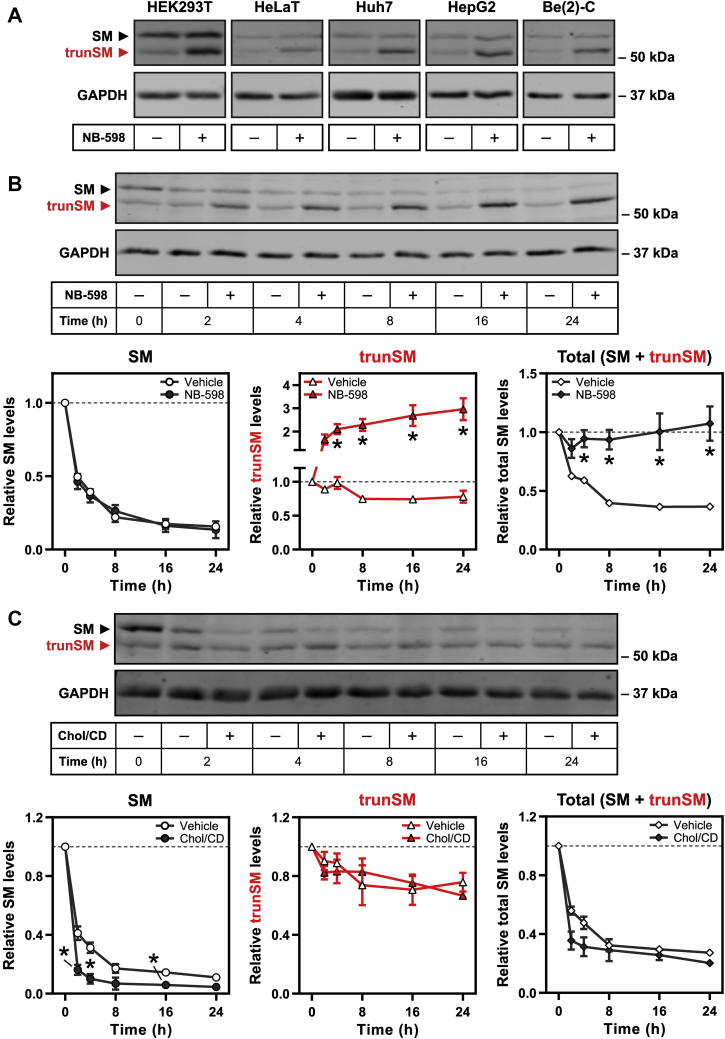
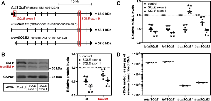
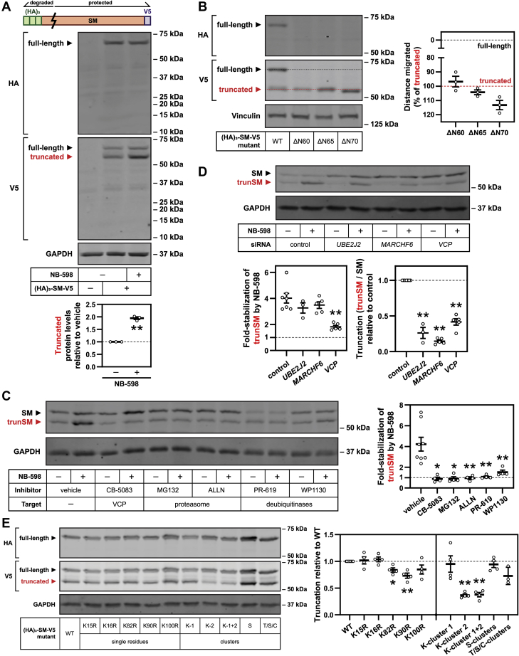
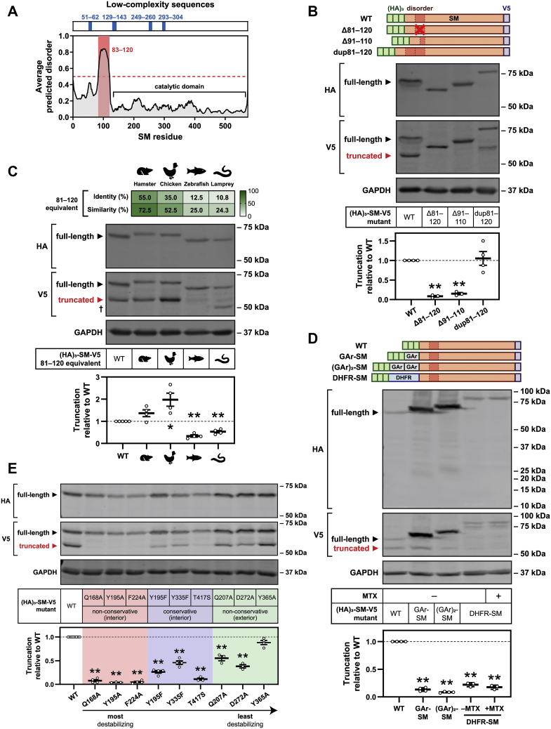
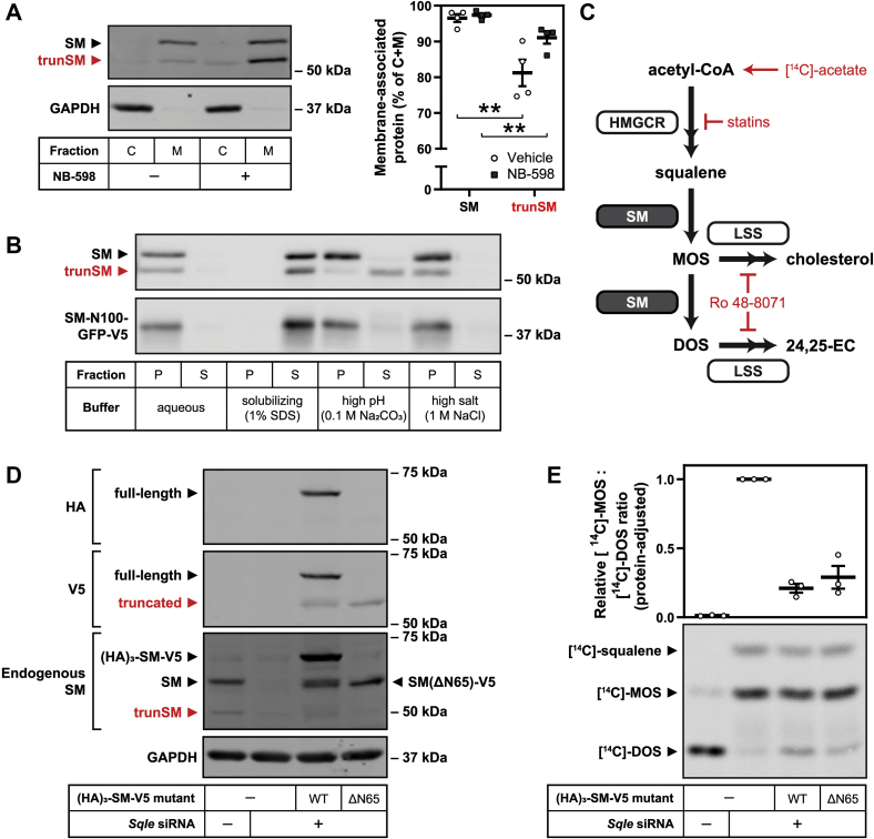
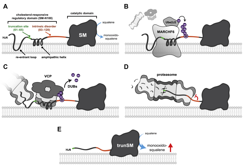
Squalene monooxygenase (SM, also known as squalene epoxidase) is a rate-limiting enzyme of cholesterol synthesis that converts squalene to monooxidosqualene and is oncogenic in numerous cancer types. SM is subject to feedback regulation via cholesterol-induced proteasomal degradation, which depends on its lipid-sensing N-terminal regulatory domain. We previously identified an endogenous truncated form of SM with a similar abundance to full-length SM, but whether this truncated form is functional or subject to the same regulatory mechanisms as full-length SM is not known. Here, we show that truncated SM differs from full-length SM in two major ways: it is cholesterol resistant and adopts a peripheral rather than integral association with the endoplasmic reticulum membrane. However, truncated SM retains full SM activity and is therefore constitutively active. Truncation of SM occurs during its endoplasmic reticulum-associated degradation and requires the proteasome, which partially degrades the SM N-terminus and disrupts cholesterol-sensing elements within the regulatory domain. Furthermore, truncation relies on a ubiquitin signal that is distinct from that required for cholesterol-induced degradation. Using mutagenesis, we demonstrate that partial proteasomal degradation of SM depends on both an intrinsically disordered region near the truncation site and the stability of the adjacent catalytic domain, which escapes degradation. These findings uncover an additional layer of complexity in the post-translational regulation of cholesterol synthesis and establish SM as the first eukaryotic enzyme found to undergo proteasomal truncation.

Keywords: cholesterol; endoplasmic reticulum–associated protein degradation; proteasome; protein degradation; squalene monooxygenase; ubiquitylation (ubiquitination).


Conflict of interest statement

Conflict of interest The authors declare that they have no conflicts of interest with the contents of this article.

Figures

Figure 1
Figure 1
A truncatedandcholesterol-resistantform of SM is present in a variety of cell types.A, the indicated cell lines were treated in the presence or the absence of 1 μM NB-598 for 8 h, and immunoblotting was performed for SM and truncated SM (trunSM; red). Immunoblots are representative of n ≥ 3 (HEK293T, HeLaT, Huh7, and HepG2) or n = 1 (Be(2)-C) independent experiments. B and C, HEK293T cells were treated with 10 μg/ml cycloheximide in the presence or the absence of (B) 1 μM NB-598 or (C) 20 μg/ml cholesterol-methyl-β-cyclodextrin complexes (Chol/CD) for the indicated times. Graphs depict densitometric quantification of SM levels (left), trunSM levels (center), or total SM levels (SM + trunSM; right) normalized to the 0 h time point, which was set to 1 (dotted line). Data presented as mean ± SEM from n ≥ 3 independent experiments (∗p ≤ 0.05; two-tailed paired t test versus vehicle condition). HEK293T, human embryonic kidney 293T; SM, squalene monooxygenase.
Figure 2
Figure 2
trunSM is not produced by alternative SQLE transcripts.A, schematic of full-length (fullSQLE) and alternative protein-coding (trunSQLE1 and trunSQLE2) SQLE transcripts. Exons and untranslated regions are indicated by black bars, and siRNA target regions are indicated by red boxes. B and C, HEK293T cells were transfected with the indicated siRNAs for 24 h and refreshed in maintenance medium for a further 24 h. B, graph depicts densitometric quantification of SM and trunSM levels normalized to the control siRNA condition, which was set to 1 (dotted line). C, SQLE transcript levels were normalized to PBGD housekeeping transcript levels and adjusted relative to the control siRNA condition, which was set to 1 (dotted line). D, absolute quantification of SQLE cDNA levels in control siRNA samples from (C). BD, data are presented as mean ± SEM from n = 4 independent experiments, each performed in triplicate for quantitative RT-PCR analysis (∗∗p ≤ 0.01; two-tailed paired t test versus control siRNA). cDNA, complementary DNA; HEK293T, human embryonic kidney 293T; SQLE, squalene epoxidase; trunSM, truncated SM.
Figure 3
Figure 3
trunSM arises from partial proteasomal degradation of the SM N-terminus.A, HEK293T cells were transfected with empty vector or (HA)3–SM–V5 expression vector for 24 h, refreshed in maintenance medium for 16 h, and treated in the presence or the absence of 1 μM NB-598 for 8 h. Lysates were separated by 4 to 15% gradient Tris–glycine SDS-PAGE. Graph depicts densitometric quantification of truncated protein levels normalized to the vehicle condition, which was set to 1 (dotted line). B, HEK293T cells were transfected with the indicated constructs for 24 h and refreshed in maintenance medium for 24 h. Lysates were separated by 7.5% Tris–glycine SDS-PAGE. Graph depicts quantification of construct migration relative to the separation of WT full-length and truncated (HA)3–SM–V5, which were set to 0% (gray dotted line) and 100% (red dotted line), respectively. C, HEK293T cells were treated with 5 μM CB-5083, 20 μM MG132, 25 μg/ml ALLN, 40 μM PR-619, or 10 μM WP1130, in the presence or the absence of 1 μM NB-598, for 8 h. Graph depicts densitometric quantification of trunSM stabilization by NB-598. D, HEK293T cells were transfected with the indicated siRNAs for 24 h, refreshed in maintenance medium for 16 h, and treated in the presence or the absence of 1 μM NB-598 for 8 h. Graphs depict densitometric quantification of (left) trunSM stabilization by NB-598 or (right) SM truncation normalized to the control siRNA condition, which was set to 1 (dotted line). E, HEK293T cells were transfected with the indicated constructs for 24 h and refreshed in maintenance medium for 24 h. Graph depicts densitometric quantification of (HA)3–SM–V5 truncation normalized to the WT construct, which was set to 1 (dotted line). Cluster mutations: K-cluster 1 (K15R and K16R); K-cluster 2 (K82R, K90R, and K100R); K-cluster 1 + 2 (K15R, K16R, K82R, K90R, and K100R); S-clusters (S59A, S61A, S83A, and S87A); T/S/C-clusters (T3A, T9A, T11A, S43A, C46A, S59A, S61A, S67A, S71A, S83A, and S87A). AE, data presented as mean ± SEM from n ≥ 3 independent experiments (∗p ≤ 0.05; ∗∗p ≤ 0.01, two-tailed paired t test versus [A and B] vehicle, [C] control siRNA, or [D] WT). HEK293T, human embryonic kidney 293T; trunSM, truncated SM.
Figure 4
Figure 4
SM truncation depends on an intrinsically disordered region and the stability of the catalytic domain.A, low-complexity regions (blue) and intrinsically disordered regions (red) within the SM protein sequence. B, C, and E, HEK293T cells were transfected with the indicated constructs for 24 h and refreshed in maintenance medium for 24 h. C, dagger indicates an additional non-trunSM fragment. D, HEK293T cells were transfected with the indicated constructs for 24 h, refreshed in maintenance medium for 16 h, and treated in the presence or the absence of 10 μM methotrexate (MTX) for 8 h. Lysates were separated by 4 to 15% gradient Tris–glycine SDS-PAGE. BE, graphs depict densitometric quantification of (HA)3–SM–V5 truncation normalized to the WT construct, which was set to 1 (dotted line). Data are presented as mean ± SEM from n ≥ 3 independent experiments (∗p ≤ 0.05; ∗∗p ≤ 0.01, two-tailed paired t test versus WT). DHFR, dihydrofolate reductase; GAr, glycine-alanine repeat; HEK293T, human embryonic kidney 293T; SM, squalene monooxygenase; trunSM, truncated SM.
Figure 5
Figure 5
trunSM has an altered ER membrane topology and retains SM activity.A, HEK293T cells were treated in the presence or the absence of 1 μM NB-598 for 8 h, and cytosolic (C) or membrane (M) fractions were isolated. Graph depicts the proportion of overall protein (C + M) found in the membrane fraction. Data are presented as mean ± SEM from n = 4 independent experiments (∗∗p ≤ 0.01, two-tailed paired t test versus SM). B, HEK293T cells were transfected with pTK–SM-N100–GFP-V5 for 24 h and refreshed in maintenance medium for a further 24 h. Membrane fractions were isolated and treated as indicated, followed by collection of pellet (P) and supernatant (S) fractions. Immunoblot is representative of n = 3 independent experiments. C, simplified schematic of the cholesterol synthesis pathway. Acetyl-CoA is converted to squalene by enzymes including HMG-CoA reductase (HMGCR), the target of statins. SM catalyzes the epoxidation of squalene to form monooxidosqualene (MOS), which is converted to cholesterol by lanosterol synthase (LSS) and other downstream enzymes. When SM activity is high or LSS activity is inhibited by Ro 48-8071, SM converts MOS to dioxidosqualene (DOS), the precursor of a shunt pathway producing 24,25-epoxycholesterol (24,25-EC). [14C]-acetate can feed into the cholesterol synthesis pathway at the level of acetyl-CoA. D, CHO-7 cells were transfected in the presence or the absence of Sqle siRNA for 24 h, refreshed in sterol-depletion medium, and transfected with the indicated constructs for 16 h. Cells were then treated with 100 nM Ro 48-8071 for 4 h. Immunoblot is representative of n = 3 independent experiments. Note that immunoblotting for endogenous SM also detects ectopic constructs. E, CHO-7 cells were treated as described in (C) and in addition labeled with [14C]-acetate during the 4 h Ro 48-8071 treatment. Cells were then assayed for the synthesis of [14C]-squalene, [14C]-MOS, or [14C]-DOS. Graph depicts quantification of [14C]-MOS:[14C]-DOS (substrate:product) ratios normalized to the condition with the highest ratio, as we have done previously (19), and adjusted for the expression of cotransfected SM constructs. Data are presented as mean ± SEM from n = 3 independent experiments. ER, endoplasmic reticulum; HEK293T, human embryonic kidney 293T; trunSM, truncated SM.
Figure 6
Figure 6
Model for the mechanism of SM truncation.A, full-length SM comprises the SM-N100 regulatory domain, containing a cholesterol-sensing re-entrant loop and amphipathic helix, and the C-terminal catalytic domain that converts squalene to monooxidosqualene. B, Ube2J2 and MARCHF6 ubiquitinate the SM-N100 domain, likely at the known ubiquitination site Lys-90. C, VCP is recruited to ubiquitinated SM and extracts the SM-N100 domain from the ER membrane, allowing the proteasome to begin degrading SM from its N-terminus. Deubiquitinases (DUBs) are required for this process. D, the SM 81 to 120 disordered region impedes the unfolding of the adjacent and highly stable catalytic domain by the proteasome, preventing further degradation. E, the undegraded portion of SM (trunSM, with a new N-terminus between residues 60 and 65) is released from the proteasome. The loss of the SM-N100 re-entrant loop and disruption of the SM-N100 amphipathic helix renders trunSM resistant to cholesterol-induced degradation and therefore constitutively active. ER, endoplasmic reticulum; SM, squalene monooxygenase; VCP, valosin-containing protein.

Similar articles

Cited by

References

    1. Ikonen E. Cellular cholesterol trafficking and compartmentalization. Nat. Rev. Mol. Cell Biol. 2008;9:125–138. - PubMed
    1. Prospective Studies Collaboration Blood cholesterol and vascular mortality by age, sex, and blood pressure: A meta-analysis of individual data from 61 prospective studies with 55 000 vascular deaths. Lancet. 2007;370:1829–1839. - PubMed
    1. Kuzu O.F., Noory M.A., Robertson G.P. The role of cholesterol in cancer. Cancer Res. 2016;76:2063–2070. - PMC - PubMed
    1. Howe V., Sharpe L.J., Alexopoulos S.J., Kunze S.V., Chua N.K., Li D., Brown A.J. Cholesterol homeostasis: How do cells sense sterol excess? Chem. Phys. Lipids. 2016;199:170–178. - PubMed
    1. Gill S., Stevenson J., Kristiana I., Brown A.J. Cholesterol-dependent degradation of squalene monooxygenase, a control point in cholesterol synthesis beyond HMG-CoA reductase. Cell Metab. 2011;13:260–273. - PubMed

Publication types