. 2021 Jan-Jun:296:100726.
doi: 10.1016/j.jbc.2021.100726. Epub 2021 Apr 30.

PIP2 regulation of TRPC5 channel activation and desensitization

Affiliations

PIP2 regulation of TRPC5 channel activation and desensitization

Mehek Ningoo et al. J Biol Chem. 2021 Jan-Jun.

Abstract

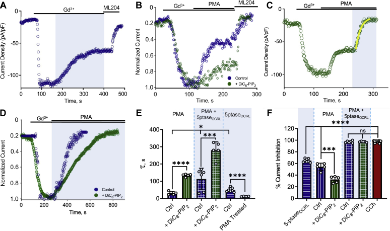
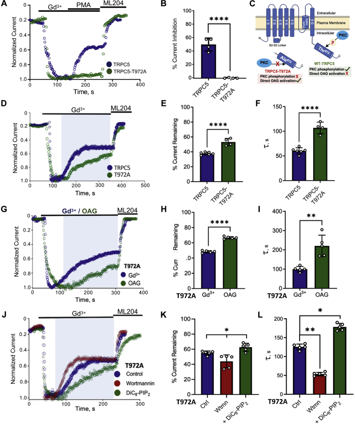
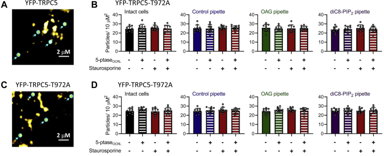
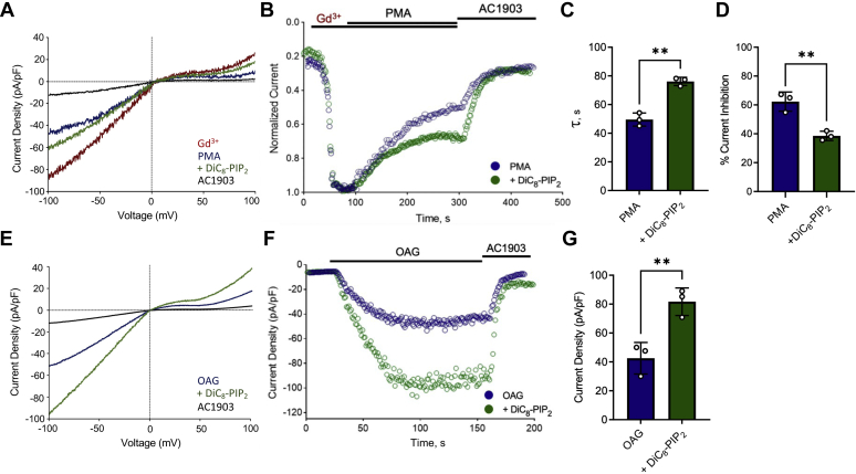
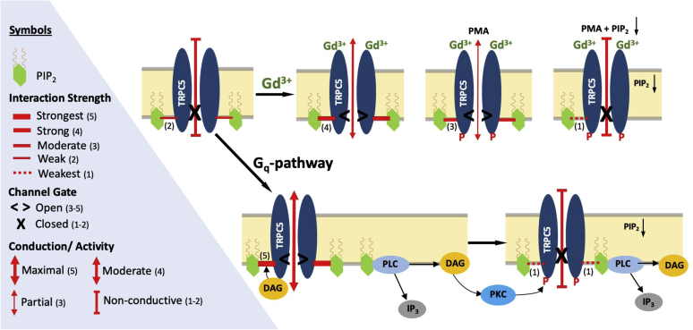
Transient receptor potential canonical type 5 (TRPC5) ion channels are expressed in the brain and kidney and have been identified as promising therapeutic targets whose selective inhibition can protect against diseases driven by a leaky kidney filter, such as focal segmental glomerular sclerosis. TRPC5 channels are activated not only by elevated levels of extracellular Ca2+or lanthanide ions but also by G protein (Gq/11) stimulation. Phosphatidylinositol 4,5-bisphosphate (PIP2) hydrolysis by phospholipase C enzymes leads to PKC-mediated phosphorylation of TRPC5 channels and their subsequent desensitization. However, the roles of PIP2 in activation and maintenance of TRPC5 channel activity via its hydrolysis product diacyl glycerol (DAG), as well as the mechanism of desensitization of TRPC5 activity by DAG-stimulated PKC activity, remain unclear. Here, we designed experiments to distinguish between the processes underlying channel activation and inhibition. Employing whole-cell patch-clamp, we used an optogenetic tool to dephosphorylate PIP2 and assess channel-PIP2 interactions influenced by activators, such as DAG, or inhibitors, such as PKC phosphorylation. Using total internal reflection microscopy, we assessed channel cell surface density. We show that PIP2 controls both the PKC-mediated inhibition and the DAG- and lanthanide-mediated activation of TRPC5 currents via control of gating rather than channel cell surface density. These mechanistic insights promise to aid in the development of more selective and precise inhibitors to block TRPC5 channel activity and illuminate new opportunities for targeted therapies for a group of chronic kidney diseases for which there is currently a great unmet need.

Keywords: TRPC5 channels; diacyl glycerol (DAG); phosphatidylinositol 4,5-bisphosphate (PIP(2)); phosphatidylinositol signaling; phosphoinositide; transient receptor potential channels (TRP channels).


Conflict of interest statement

Conflict of interest A. G. has a financial interest in Goldfinch Biopharma, which was reviewed and is managed by Brigham and Women’s Hospital, Mass General Brigham (MGB), and the Broad Institute of MIT and Harvard in accordance with their conflict-of-interest policies. All other authors declare that they have no conflicts of interest with the contents of this article.

Figures

Figure 1
Figure 1
PIP2implicated in PKC-mediated desensitization and promotion of Gd3+-activated TRPC5 currents.A, left, example current density and voltage relationships for HEK293T cells expressing mTRPC5–GFP. Currents were evoked by a ramp from −100 mV to 100 mV followed by application of 100 μM CCh. Cells were studied under control conditions (blue) or with PIP-5K coexpression (green) or with 200 μM diC8–PIP2 in the pipette (purple). The spontaneous decrease in current is illustrated by sweeps labeled 1 to 5, which correspond to 50-s intervals, as illustrated on the exemplar time courses (right). B, the mean of percentage current remaining during 100 μM CCh treatment in control HEK293T cells expressing mTRPC5–GFP (n = 12, 0.89 ± 2.948), with overexpression of PIP-5K (n = 9, 47.07 ± 5.78) and with diC8–PIP2 in the pipette (n = 10, 52.32 ± 5.89). C, the bar graph of time taken from peak to 50% current decay (T50) of control HEK293T cells expressing mTRPC5–GFP (n = 11, 68.82 ± 8.681) and after treatment with 20 μM wortmannin for 1 h (n = 9, 10.23 ± 1.722); inset: representative whole-cell patch-clamp recordings in each condition of HEK293T cells expressing TRPC5–GFP activated by 100 μM CCh. D, current density voltage curves of the ±100 mV ramp of ML204-sensitive currents activated by 100 μM Gd3+ activation in TRPC5–GFP expressing HEK293T cells (control) and after 1 h treatment with 20 μM wortmannin. E, representative whole-cell current density (pA/pF) curves observed in HEK293T cells overexpressing mTRPC5–GFP activated with 100 μM GdCl3 and upon 20 μM wortmannin treatment for 1 h. F, the bar graph of Ip (peak current density-pA/pF) of control HEK293T cells expressing TRPC5–GFP (n = 5, 191 ± 25.23) and cells treated with wortmannin (n = 5,49.35 ± 6.5). Values reported as mean ± SD, p-values established using Student’s t test, ∗∗∗∗p < 0.0001. CCh, carbachol; diC8–PIP2, dioctanoyl-glycerol-PIP2; PIP-5K, phosphatidylinositol 4-phosphate 5 kinase; TRPC5, transient receptor potential canonical type 5.
Figure 2
Figure 2
TRPC5 current inhibition by PKC-mediated phosphorylation and/or PIP2dephosphorylation reveals an underlying decrease in channel–PIP2interactions.A, whole-cell patch clamp recording of HEK293T cells expressing TRPC5–GFP, light-activated CRY2–5’PTASEOCRL, and CIBN–CAAX–GFP (see Experimental procedures); inward current activated by 100 μM GdCl3 with channel current decrease in response to light-activated metabolism of PIP2 and remaining current blocked by 3 μM ML204. B, inhibition observed by PKC activator PMA without/with 200 μM diC8–PIP2 in the pipette. C, HEK-293T cells expressing TRPC5–GFP, CRY2–5’ptase, and CIBN–CAAX–GFP were activated using 100 μM GdCl3; 200 nM PMA was applied to activate PKC enzymes followed by blue-light exposure. D, inhibition observed by simultaneous application of PKC activator PMA and activation of light-activated inositol phosphatase without/with 200 μM diC8–PIP2 in the pipette. E, the bar graph of the mean decay constant ± SD of PMA-mediated inhibition alone (n = 5, 31.44 ± 9.62) and with diC8–PIP2 (n = 5, 133.95 ± 7.766), simultaneous PMA and 5’-ptaseOCRL–mediated inhibition (n = 6, 112.97 ± 62.87) and with diC8–PIP2 (n = 5, 280.33 ± 48.71), and 5’-ptaseOCRL-mediated inhibition alone (n = 8, 52.57 ± 11.59) and after PMA treatment (n = 5, 11.43 ± 1.834). F, the bar graph summary of the mean percentage current inhibition ± SD by 5’-ptaseOCRL (n = 8, 65.18 ± 3.046), PMA-mediated inhibition alone (n = 5, 54.4 ± 7.23), and with diC8–PIP2 (n = 5, 32.47 ± 6.04), simultaneous PMA and 5’-ptaseOCRL-mediated inhibition (n = 6, 97.51 ± 2.397) and with diC8–PIP2 (n = 5, 96.87 ± 0.8152), and when activated using 100 μM CCh (n = 12, 97.61 ± 2.32). Values reported as mean ± SD, p-values established using Students’ t test, comparison with experimental control (#); ∗p < 0.05, ∗∗∗p < 0.001, ∗∗∗∗p < 0.0001. CCh, carbachol; CRY2, cryptochrome 2; diC8–PIP2, dioctanoyl-glycerol-PIP2; PMA, phorbol 12-myristate-13-acetate; TRPC5, transient receptor potential canonical type 5.
Figure 3
Figure 3
OAG-mediated activation of TRPC5 channels shows enhanced channel–PIP2interaction strength.A, HEK-293T cells expressing TRPC5–GFP/TRPC5–T972A–GFP were activated using 100 μM GdCl3, and effect of 200 nM PMA was observed. B, the bar graph summary of the mean percentage current inhibition by PMA in WT TRPC5 (n = 4, 50.18 ± 9.87) and PKC-insensitive TRPC5–T972A mutant (n =4, 0.75 ± 0.96). C, cartoon depicting the differences between the WT and PKC-insensitive TRPC5–T972A mutant channels. D, HEK-293T cells expressing TRPC5–GFP/TRPC5–T972A–GFP, CRY2–5’ptase, and CIBN–CAAX–GFP were activated using 100 μM GdCl3 and the effect of blue-light exposure was observed. E, the bar graph summary of the mean percentage current remaining ± SD (in panel D) in WT TRPC5 (n = 6, 37.83 ± 1.8392) and PKC-insensitive TRPC5–T972A mutant (n = 5, 53.175 ± 4.39). F, the bar graph of the mean decay constant of inhibition ± SD (in panel D) for TRPC5 (n = 6, 60.27 ± 9.79) for mTRPC5–T972A (n = 4, 106.67 ± 14.42). G, HEK-293T cells expressing TRPC5–T972A–GFP, CRY2–5’ptase, and CIBN–CAAX–GFP were activated using saturated concentration of Gd3+ (150 μM) or OAG (200 μM), and the effect of blue-light exposure was observed. H, the bar graph summary of the mean percentage current remaining ± SD (in panel G) in TRPC5–T972A upon activation by Gd3+ (n = 5, 48.32 ± 1.45) and OAG (n = 5, 66.7 ± 1.51). I, the bar graph of the mean decay constant of inhibition ± SD (in panel G) when activated by 150 μM Gd3+ (n = 5, 99.55 ± 14.11) and 200 μM of OAG (n = 5, 220.5 ± 56.03). J, HEK-293T cells expressing TRPC5–T972A–GFP, CRY2–5’ptase, and CIBN–CAAX–GFP were activated using 100 μM OAG (control) and incubated in 20 μM wortmannin for 1 h and 200 μM diC8–PIP2 in the pipette. K, the bar graph summary of the mean percentage current remaining ± SD (in panel J) in TRPC5–T972A upon activation by OAG (n = 5, 54.84 ± 7.51), with wortmannin (n =5, 43.94 ± 8.6) and diC8–PIP2 (n = 5, 62.64 ± 5.3). L, the bar graph of the mean decay constant of inhibition ± SD (in panel J) for control (n = 5, 125.5 ± 7.5), with wortmannin (n = 5, 53.25 ± 4.65) and with diC8–PIP2 (n = 5, 178.25 ± 9.45). Values reported as mean ± SD, p-values established using Students’ t test, ∗p < 0.05, ∗∗p < 0.01, ∗∗∗∗p < 0.0001. CRY2, cryptochrome 2; diC8–PIP2, dioctanoyl-glycerol-PIP2; PMA, phorbol 12-myristate-13-acetate; TRPC5, transient receptor potential canonical type 5.
Figure 4
Figure 4
Regulation by OAG or PIP2does not alter the surface density of YFP–TRPC5 channels. YFP-tagged mTRPC5 or mTRPC5–T972A channels were expressed in HEK293T cells and studied by patch clamp TIRF. The number of fluorescent particles was determined 200 s after whole-cell mode was established to allow dialysis of the cells with the control solution (blue), 200 μM OAG (green) or 200 μM diC8–PIP2 (purple). Cells were studied with or without optogenetic activation of 5’-ptaseOCRL (white stripped bars) or after incubation with 1 μM staurosporine (red). Bar graphs represent particle density as the mean ± SD number of fluorescent particles in the TIRF field in 3 to 6 random 10 × 10 μm squares per cell and from 4 to 6 cells per group. A, TIRF image showing YFP-tagged WT mTRPC5 channels at the cell surface. Four example particles corresponding to single TRPC5 channels are highlighted in cyan. B, the bar graphs summarizing the density of fluorescent particles indicating no change from control values under any of the conditions studied. C, TIRF image showing YFP-tagged mTRPC5–T972A channels at the cell surface. Four example particles corresponding to single TRPC5 channels are highlighted in cyan. D, the bar graphs summarizing the density of fluorescent particles indicating no change from control values under any of the conditions studied. Values reported as mean ± SD, p-values established using Students’ t test. diC8–PIP2, dioctanoyl-glycerol-PIP2; TRPC5, transient receptor potential canonical type 5.
Figure 5
Figure 5
PIP2prevents PKC-mediated desensitization and promotes OAG-mediated activation in endogenously expressed TRPC5 channels. The experiments shown in this figure were carried out in HT-22 murine cells that predominantly express TRPC5 channels (2). A, current density voltage curves of the ± 100 mV ramp of 100 μM Gd3+, PMA inhibition with/without 200 μM diC8–PIP2 in the pipette and inhibition with 100 μM AC1903 (a small-molecule selective inhibitor of TRPC5). B, representative whole-cell recording of PMA-mediated inhibition of Gd3+ with/without 200 μM diC8–PIP2.C, the bar graph summary of the mean decay constant of inhibition observed with PMA (control n = 3, 49.5 ± 4.55) and with 200 μM diC8–PIP2 (n = 3, 76 ± 2.87). D, the bar graph summary of mean percentage current inhibited ± SD with PMA (control n = 3, 62.26 ± 6.37) and with 200 μM diC8–PIP2 (n = 3, 38.8 ± 3.23). E, current density voltage curves of the ±100 mV ramp in HT-22 cells treated with 1 μM staurosporine for 30 min, of 100 μM OAG activation with/without 200 μM diC8–PIP2 in the pipette, and inhibition with 100 μM AC1903. F, representative whole-cell recording of 100 μM OAG-activated currents after treatment with 1 μM staurosporine for 30 min, with/without 200 μM diC8–PIP2.G, the bar graph summary of mean peak current density ± SD observed with 100 μM OAG (control n = 3, 42.5 ± 6.305) and with 200 μM diC8–PIP2 (n = 3, 81.6 ± 5.51). Values reported as mean ± SD, p-values established using Students' t-test ∗∗p < 0.01. diC8–PIP2, dioctanoyl-glycerol-PIP2; PMA, phorbol 12-myristate-13-acetate; TRPC5, transient receptor potential canonical type 5.
Figure 6
Figure 6
Cartoon model of the dependency of TRPC5 channel on PIP2to account for stimulation and inhibition of channel activity by independent gating mechanisms. Trivalent cation-mediated control of TRPC5 activity: Trivalent cation activation mediated by Gd3+ allosterically strengthens channel interactions with PIP2, strongly enough to cause partial activation. PMA treatment alone (PKC-mediated phosphorylation but not PIP2 depletion) weakens channel–PIP2 interaction strength and causes partial inhibition of channel currents. Similarly, depletion of intracellular PIP2 levels (using either wortmannin or 5’-phosphatase) alone (PIP2 depletion but not PKC-mediated phosphorylation) does not strip the channel completely of its PIP2 causing partial inhibition of activity. The combination of PMA treatment and PIP2 depletion strips the channel from its PIP2 severely enough to cause full inhibition. Gq-mediated control of TRPC5 activity: upon Gq-receptor activation, PLC hydrolyzes PIP2 to IP3 and DAG. DAG allosterically enhances stronger channel interactions with PIP2, activating the channel maximally. PIP2 depletion (such as by dephosphorylation of PIP2) alone (without PKC-mediated phosphorylation as in T972A) causes partial inhibition. The ensuing DAG activation of PKC causes channel phosphorylation at T972, which allosterically weakens channel–PIP2 interactions enough that adds up to the PIP2 depletion causing full inhibition of the current. IP3, inositol 1,4,5-triphosphate; PLC, phospholipase C; PMA, phorbol 12-myristate-13-acetate; TRPC5, transient receptor potential canonical type 5.

References

    1. Clapham D.E. TRP channels as cellular sensors. Nature. 2003;426:517–524. - PubMed
    1. Storch U., Forst A.L., Pardatscher F., Erdogmus S., Philipp M., Gregoritza M., y Schnitzler M.M., Gudermann T. Dynamic NHERF interaction with TRPC4/5 proteins is required for channel gating by diacylglycerol. Proc. Natl. Acad. Sci. U. S. A. 2017;114:37–46. - PMC - PubMed
    1. Schaldecker T., Kim S., Tarabanis C., Tian D., Hakroush S., Castonguay P., Ahn W., Wallentin H., Heid H., Hopkins C.R., Lindsley C.W., Riccio A., Buvall L., Weins A., Greka A. Inhibition of the TRPC5 ion channel protects the kidney filter. J. Clin. Invest. 2013;123:5298–5309. - PMC - PubMed
    1. Bezzerides V.J., Ramsey I.S., Kotecha S., Greka A., Clapham D.E. Rapid vesicular translocation and insertion of TRP channels. Nat. Cell Biol. 2004;6:709–720. - PubMed
    1. Trebak M., Lemonnier L., DeHaven W.I., Wedel B.J., Bird G.S., Putney J.W. Complex functions of phosphatidylinositol 4, 5-bisphosphate in regulation of TRPC5 cation channels. Pflügers Arch. 2009;457:757–769. - PMC - PubMed

Publication types

Substances

LinkOut - more resources