

Parameters Indicating Development of Influenza-Associated Acute Necrotizing Encephalopathy: Experiences from a Single Center
- PMID: 33934098
- PMCID: PMC8101270
- DOI: 10.12659/MSM.930688
Parameters Indicating Development of Influenza-Associated Acute Necrotizing Encephalopathy: Experiences from a Single Center
Abstract
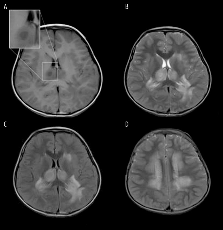
BACKGROUND Influenza-associated acute necrotizing encephalopathy (IANE) can be lethal and disabling and have a sudden onset and deteriorate rapidly but lacks early diagnostic indicators. We aimed to examine the early clinical diagnostic indicators in children with IANE. MATERIAL AND METHODS Acute influenza patients were grouped according to their clinical manifestations: flu alone (FA), flu with febrile seizure (FS), influenza-associated encephalopathy (IAE), and IANE. The clinical features, biomarkers, neuroelectrophysiological results, and neuroimaging examination results were compared. RESULTS A total of 31 patients were included (FA (n=4), FS (n=8), IAE (n=14), and IANE (n=5)). The IANE group, whose mean age was 3.7 years, was more likely to show rapid-onset seizure, acute disturbance of consciousness (ADOC), Babinski's sign, and death/sequela. More patients in the IANE group required tracheal intubation mechanical ventilation and received intravenous immunoglobulins (IVIG) and glucocorticoids. The alanine aminotransferase (ALT), aspartate transaminase (AST), and lactate dehydrogenase (LDH) levels in the IANE group were significantly higher than in the FS and IAE groups. The aquaporin-4 (AQP-4) antibody and malondialdehyde (MDA) levels in the serum and cerebrospinal fluid (CSF) were notably higher in IANE patients in the acute stage compared with FS and IAE patients. All patients in the IANE group had positive neuroimaging findings. CONCLUSIONS Early clinical warning factors for IANE include rapid-onset seizures in patients under 4 years of age, ADOC, and pathological signs. Increased AQP-4 antibodies and MDA levels in CSF might contribute to early diagnosis. Early magnetic resonance venography (MRV) and susceptibility-weighted imaging (SWI) sequences, or thrombelastography to identify deep vein thrombosis, might indicate clinical deterioration.
Conflict of interest statement
None.
Figures


References
-
- Shiomi M. [Pathogenesis of acute encephalitis and acute encephalopathy]. Nihon Rinsho. 2011;69:399–408. [in Japanese] - PubMed
-
- Okumura A, Abe S, Kidokoro H, et al. Acute necrotizing encephalopathy: A comparison between influenza and non-influenza cases. Microbiol Immunol. 2009;53:277–80. - PubMed
-
- Kondrich J, Rosenthal M. Influenza in children. Curr Opin Pediatr. 2017;29:297–302. - PubMed
-
- Shah S, Keil A, Gara K, et al. Neurologic complications of influenza. J Child Neurol. 2014;29:NP49-53. - PubMed
MeSH terms
Substances
LinkOut - more resources
Full Text Sources
Other Literature Sources
Medical
