

Cortical spectral matching and shape and volume analysis of the fetal brain pre- and post-fetal surgery for spina bifida: a retrospective study
- PMID: 33934181
- PMCID: PMC8460513
- DOI: 10.1007/s00234-021-02725-8
Cortical spectral matching and shape and volume analysis of the fetal brain pre- and post-fetal surgery for spina bifida: a retrospective study
Abstract
Purpose: A retrospective study was performed to study the effect of fetal surgery on brain development measured by MRI in fetuses with myelomeningocele (MMC).
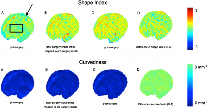
Methods: MRI scans of 12 MMC fetuses before and after surgery were compared to 24 age-matched controls without central nervous system abnormalities. An automated super-resolution reconstruction technique generated isotropic brain volumes to mitigate 2D MRI fetal motion artefact. Unmyelinated white matter, cerebellum and ventricles were automatically segmented, and cerebral volume, shape and cortical folding were thereafter quantified. Biometric measures were calculated for cerebellar herniation level (CHL), clivus-supraocciput angle (CSO), transverse cerebellar diameter (TCD) and ventricular width (VW). Shape index (SI), a mathematical marker of gyrification, was derived. We compared cerebral volume, surface area and SI before and after MMC fetal surgery versus controls. We additionally identified any relationship between these outcomes and biometric measurements.
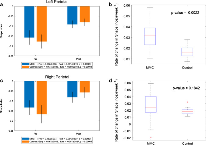
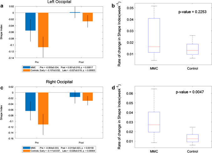
Results: MMC ventricular volume/week (mm3/week) increased after fetal surgery (median: 3699, interquartile range (IQR): 1651-5395) compared to controls (median: 648, IQR: 371-896); P = 0.015. The MMC SI is higher pre-operatively in all cerebral lobes in comparison to that in controls. Change in SI/week in MMC fetuses was higher in the left temporal lobe (median: 0.039, IQR: 0.021-0.054), left parietal lobe (median: 0.032, IQR: 0.023-0.039) and right occipital lobe (median: 0.027, IQR: 0.019-0.040) versus controls (P = 0.002 to 0.005). Ventricular volume (mm3) and VW (mm) (r = 0.64), cerebellar volume and TCD (r = 0.56) were moderately correlated.
Conclusions: Following fetal myelomeningocele repair, brain volume, shape and SI were significantly different from normal in most cerebral layers. Morphological brain changes after fetal surgery are not limited to hindbrain herniation reversal. These findings may have neurocognitive outcome implications and require further evaluation.
Keywords: Cortical spectral matching; Fetal surgery; MRI; Myelomeningocele; Shape; Volume.
© 2021. The Author(s).
Conflict of interest statement
The authors have no relevant financial or non-financial interests to disclose.
Figures


Similar articles
-
Assessment of longitudinal brain development using super-resolution magnetic resonance imaging following fetal surgery for open spina bifida.Ultrasound Obstet Gynecol. 2023 Nov;62(5):707-720. doi: 10.1002/uog.26244. Ultrasound Obstet Gynecol. 2023. PMID: 37161647 Free PMC article.
-
First 60 fetal in-utero myelomeningocele repairs at Saint Louis Fetal Care Institute in the post-MOMS trial era: hydrocephalus treatment outcomes (endoscopic third ventriculostomy versus ventriculo-peritoneal shunt).Childs Nerv Syst. 2017 Jul;33(7):1157-1168. doi: 10.1007/s00381-017-3428-8. Epub 2017 May 3. Childs Nerv Syst. 2017. PMID: 28470384
-
Comparison of brain microstructure after prenatal spina bifida repair by either laparotomy-assisted fetoscopic or open approach.Ultrasound Obstet Gynecol. 2020 Jan;55(1):87-95. doi: 10.1002/uog.20373. Ultrasound Obstet Gynecol. 2020. PMID: 31219638
-
In utero repair of spina bifida.Am J Perinatol. 2014 Aug;31(7):595-604. doi: 10.1055/s-0034-1372429. Epub 2014 May 12. Am J Perinatol. 2014. PMID: 24819146 Review.
-
Fetal surgery for spina bifida: past, present, future.Semin Pediatr Surg. 2013 Feb;22(1):10-7. doi: 10.1053/j.sempedsurg.2012.10.003. Semin Pediatr Surg. 2013. PMID: 23395140 Free PMC article. Review.
Cited by
-
The Role of Fetal Brain Magnetic Resonance Imaging in Current Fetal Medicine.J Belg Soc Radiol. 2022 Dec 13;106(1):130. doi: 10.5334/jbsr.3000. eCollection 2022. J Belg Soc Radiol. 2022. PMID: 36569393 Free PMC article.
-
Semi-automatic segmentation of the fetal brain from magnetic resonance imaging.Front Neurosci. 2022 Nov 11;16:1027084. doi: 10.3389/fnins.2022.1027084. eCollection 2022. Front Neurosci. 2022. PMID: 36440277 Free PMC article.
-
Thalamic connectivity topography in newborns with spina bifida: association with neurological functional level but not developmental outcome at 2 years.Cereb Cortex. 2024 Jan 14;34(1):bhad438. doi: 10.1093/cercor/bhad438. Cereb Cortex. 2024. PMID: 37991274 Free PMC article.
-
A spatio-temporal atlas of the developing fetal brain with spina bifida aperta.Open Res Eur. 2022 Aug 31;1:123. doi: 10.12688/openreseurope.13914.2. eCollection 2021. Open Res Eur. 2022. PMID: 37645096 Free PMC article.
-
Assessment of longitudinal brain development using super-resolution magnetic resonance imaging following fetal surgery for open spina bifida.Ultrasound Obstet Gynecol. 2023 Nov;62(5):707-720. doi: 10.1002/uog.26244. Ultrasound Obstet Gynecol. 2023. PMID: 37161647 Free PMC article.
References
-
- Zarutskie A, Guimaraes C, Yepez M, Torres P, Shetty A, Sangi-Haghpeykar H, Lee W, Espinoza J, Shamshirsaz AA, Nassr A, Belfort MA, Whitehead WE, Sanz Cortes M. Prenatal brain imaging for predicting need for postnatal hydrocephalus treatment in fetuses that had neural tube defect repair in utero. Ultrasound Obstet Gynecol. 2019;53(3):324–334. doi: 10.1002/uog.20212. - DOI - PubMed
MeSH terms
Grants and funding
LinkOut - more resources
Full Text Sources
Other Literature Sources
Medical
