Review
. 2021 Jul;226(6):1659-1676.
doi: 10.1007/s00429-021-02284-x. Epub 2021 May 2.

Auditory thalamus dysfunction and pathophysiology in tinnitus: a predictive network hypothesis

Affiliations
Review

Auditory thalamus dysfunction and pathophysiology in tinnitus: a predictive network hypothesis

Pia Brinkmann et al. Brain Struct Funct. 2021 Jul.

Abstract

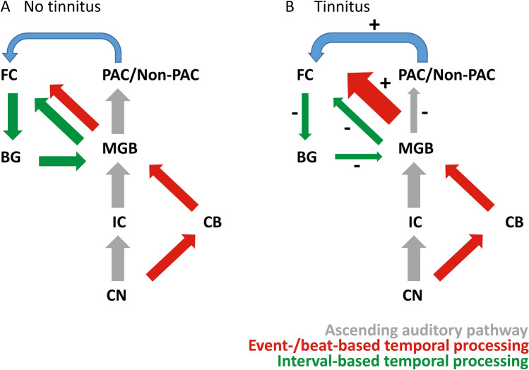
Tinnitus is the perception of a 'ringing' sound without an acoustic source. It is generally accepted that tinnitus develops after peripheral hearing loss and is associated with altered auditory processing. The thalamus is a crucial relay in the underlying pathways that actively shapes processing of auditory signals before the respective information reaches the cerebral cortex. Here, we review animal and human evidence to define thalamic function in tinnitus. Overall increased spontaneous firing patterns and altered coherence between the thalamic medial geniculate body (MGB) and auditory cortices is observed in animal models of tinnitus. It is likely that the functional connectivity between the MGB and primary and secondary auditory cortices is reduced in humans. Conversely, there are indications for increased connectivity between the MGB and several areas in the cingulate cortex and posterior cerebellar regions, as well as variability in connectivity between the MGB and frontal areas regarding laterality and orientation in the inferior, medial and superior frontal gyrus. We suggest that these changes affect adaptive sensory gating of temporal and spectral sound features along the auditory pathway, reflecting dysfunction in an extensive thalamo-cortical network implicated in predictive temporal adaptation to the auditory environment. Modulation of temporal characteristics of input signals might hence factor into a thalamo-cortical dysrhythmia profile of tinnitus, but could ultimately also establish new directions for treatment options for persons with tinnitus.

Keywords: MGB; Medial geniculate nucleus; Prediction; Temporal processing; Tinnitus.


Conflict of interest statement

The authors declare that they have no conflicts of interest.

Figures

Fig. 1
Fig. 1
Schematic and simplified representation of the classical and non-classical ascending auditory pathway. Ascending auditory signal travels from the ears to primary and secondary auditory cortices, while taking two different pathways. PAC primary auditory cortex, Non-PAC non-primary auditory cortices, CN cochlear nucleus, ICC central inferior colliculus, ICD dorsal inferior colliculus, ICX, external inferior colliculus, MGB medial geniculate body, MGD dorsal MGB, MGM medial MGB, MGV ventral MGB
Fig. 2
Fig. 2
Summary and schematic representation of increased/decreased functional connectivity measures between the MGB and cortical areas. The representation is based on baseline measures of Lv et al. (2020), Berlot et al. (2020), and Zhang et al. (2015). Depicted are only areas with altered connections to the bilateral MGB. Zhang et al. (2015) observed decreased connectivity between the left thalamus to the medial frontal gyrus and the right thalamus and superior frontal gyrus, contrasting with increased connectivity between the MGB and the IFG (BA 45) by Lv et al. (2020). Zhang et al. (2015) further observed increased connectivity between the left thalamus and the middle cingulate cortex, and the right thalamus and the posterior cingulate cortex. ACC, Anterior cingulate cortex (BA 33), PAC, Primary auditory cortex, Non-PAC, Non-Primary auditory cortices, IFG, Inferior frontal gyrus, MGB, Medial geniculate body, PostCB, Posterior cerebellum. Lv et al. (2020), Berlot et al. (2020), Zhang et al. (2015)
Fig. 3
Fig. 3
Schematic representation of the neural architecture for specific temporal prediction in persons without tinnitus a and with tinnitus b. Here, the ascending auditory pathway does not distinguish between the classical and the non-classical auditory pathway. The schema does not depict predictive top–down modulation of the network by dynamic input. The MGB forms a major hub in transmitting a timing signal to higher cortical areas (event-/beat-based temporal processing (red)). This signal forms the basis for interval-based temporal processing (green) in BG circuits. Parallel activation and integration of memory representations recruit connections between temporal and frontal cortices (blue). In tinnitus (B), connections between the MGB and auditory cortices are reduced. Starting from the MGB, increased burst and spontaneous firing leads to an increase in event-/beat-based temporal processing. Tonic firing is proposed to be reduced, reflected by decreased interval-based temporal processing, as depicted by the different arrow sizes + and – signs. In severe deafferentiation, memory retrieval increasingly relies on parahippocampal and auditory areas. PAC/Non-PAC primary and non-primary auditory cortices, BG basal ganglia, CB cerebellum, CN cochlear nucleus, FC frontal cortex, IC inferior colliculus, MGB medial geniculate body

References

    1. Aitkin L. The auditory midbrain: structure and function in the central auditory pathway. Springer; 1986.
    1. Aitkin LM, Webster WR. Medial geniculate body of the cat: organization and responses to tonal stimuli of neurons in ventral division. J Neurophysiol. 1972;35:365–380. doi: 10.1152/jn.1972.35.3.365. - DOI - PubMed
    1. Aldhafeeri FM, Mackenzie I, Kay T, Alghamdi J, Sluming V. Neuroanatomical correlates of tinnitus revealed by cortical thickness analysis and diffusion tensor imaging. Neuroradiology. 2012;54:883–892. doi: 10.1007/s00234-012-1044-6. - DOI - PubMed
    1. Allan TW, Besle J, Langers DR, Davies J, Hall DA, Palmer AR, Adjamian P. Neuroanatomical alterations in tinnitus assessed with magnetic resonance imaging. Front Aging Neurosci. 2016;8:221. doi: 10.3389/fnagi.2016.00221. - DOI - PMC - PubMed
    1. Allman MJ, Meck WH. Pathophysiological distortions in time perception and timed performance. Brain. 2012;135:656–677. doi: 10.1093/brain/awr210. - DOI - PMC - PubMed

LinkOut - more resources