. 2021 May 12;13(18):21007-21017.
doi: 10.1021/acsami.1c03288. Epub 2021 May 3.

Carbon Nanotubes/Regenerated Silk Composite as a Three-Dimensional Printable Bio-Adhesive Ink with Self-Powering Properties

Affiliations

Carbon Nanotubes/Regenerated Silk Composite as a Three-Dimensional Printable Bio-Adhesive Ink with Self-Powering Properties

Silvia Bittolo Bon et al. ACS Appl Mater Interfaces. .

Abstract

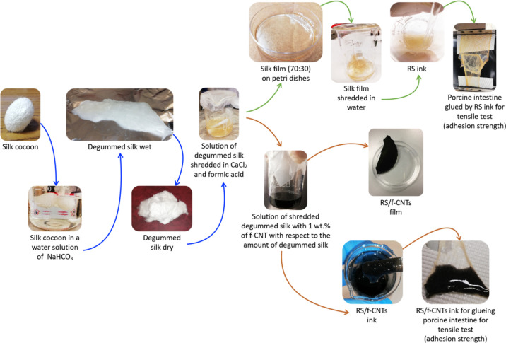
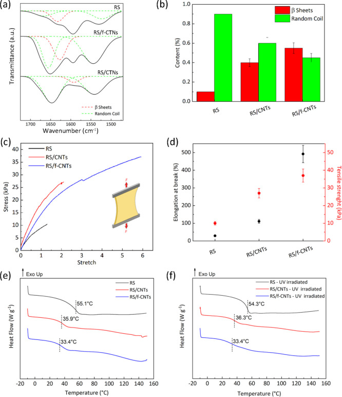
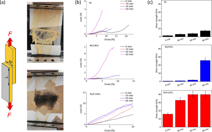
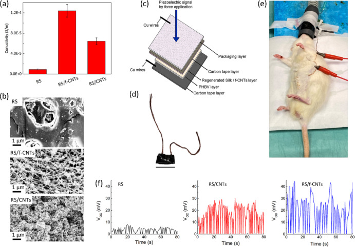
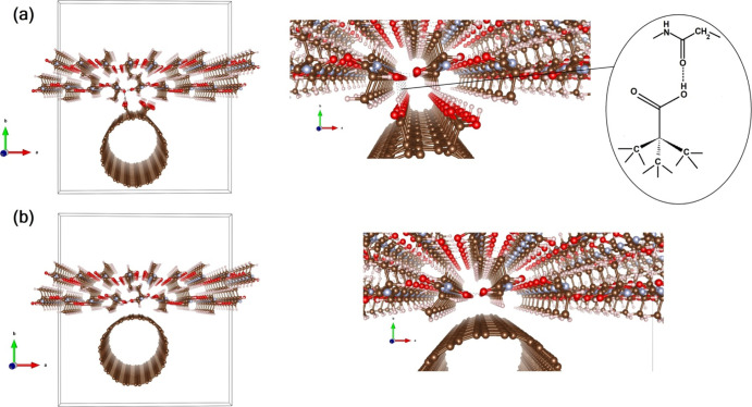
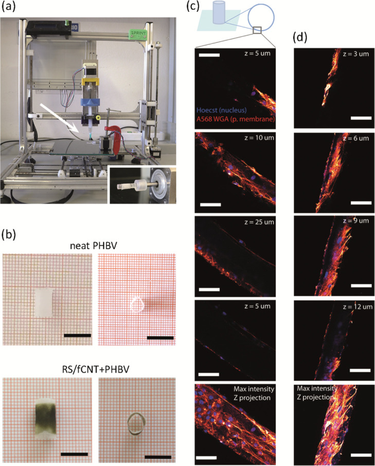
In this study, regenerated silk (RS) obtained from Bombyx Mori cocoons is compounded with carboxyl-functionalized carbon nanotubes (f-CNTs) in an aqueous environment for the fabrication of functional bio-adhesives. Molecular interactions between RS and carboxyl groups of CNTs result in structural increase of the β-sheet formation, obtaining a resistant adhesive suitable for a wet biological substrate. Moreover, the functionalization of CNTs promotes their dispersion in RS, thus enabling the production of films with controlled electrical conductivity. The practical utility of such a property is demonstrated through the fabrication of a piezoelectric device implanted in a rat to monitor the breathing in vivo and to be used as a self-powered system. Finally, RS/f-CNTs were used as a printable biomaterial ink to three dimensionally print bilayer hollow tubular structures composed of poly(3-hydroxybutyrate-co-3-hydroxyvalerate) (PHBV) and RS. Initial tests carried out by seeding and growing human skin fibroblasts demonstrated that the 3D printed bilayer hollow cylindrical structures offer a suitable surface for the seeded cells to attach and proliferate. In general, the herein proposed RS/f-CNT composite serves as a versatile material for solvent-free dispersion processing and 3D printing, thus paving a new approach to prepare multifunctional materials with potential applications of great interest in sealing biological substrates and implantable devices for regenerative medicine.

Keywords: 3D printing; carbon nanotubes; interface modeling; mechanical properties; regenerated silk; self-powering bio-adhesives.


Conflict of interest statement

The authors declare no competing financial interest.

Figures

Figure 1
Figure 1
Schematic illustration of the solvent-free fabrication method of RS/f-CNT dispersion. Silk cocoons were degummed and solubilized in FA and left to evaporate. RS was then re-dispersed in water by adding f-CNTs followed by sonication; the RS/f-CNTs can take different consistencies from ink to thin solid films characterized by high flexibility.
Figure 2
Figure 2
(a) FTIR spectra in amide-I and amide II regions of RS, RS/f-CNTs, and RS/CNTs. Peak deconvolution carried out by peak assignment to the secondary structures: 1650 cm–1 (random coil) and 1620 cm–1 (β-sheet) for amide I and 1540 cm–1 (random coil) for amide II. (b) Quantitative analysis of secondary structures in RS, RS/CNTs, and RS/f-CNTs films, respectively. (c) Stress/stretch curves for the RS, RS/CNTs, and RS/f-CNTs films. (d) Tensile strength and elongation at break of the prepared specimens calculated from tensile curves. DSC thermograms for the RS, RS/CNTs, and RS/f-CNTs films (e) before and (f) after UV irradiation, respectively. Dashed lines indicate the Tg values.
Figure 3
Figure 3
(a) Setup for measurement of shear strength (F, force; L, length; W, width). The photographs show RS (top) and RS/f-CNTs (bottom) inks adhered on porcine intestine. (b) Lap-shear tests and (c) shear strengths between porcine intestine of RS, RS/CNTs, and RS/f-CNTs adhesives as a function of the elapsed time from the deposition storing the samples at 37 °C and relative humidity of 65%.
Figure 4
Figure 4
(a) Electrical conductivity of RS, RS/CNTs, and RS/f-CNTs films, respectively. (b) FESEM images of the surface morphologies of RS, RS/f-CNTs, and RS/CNTs films, respectively. (c) Schematic representation of the sensor layout and (d) Photograph of the fabricated piezoelectric device (scale bar indicates 1 cm). (e) Photograph illustrating the implanted sensor implanted sensor in a subcutaneous pocket of the rat abdomen. (f) Open-circuit voltage (VOC) data recorded by RS, RS/CNTs, and RS/f-CNTs films, showing the signal generated by the breathing of the rat under anesthesia.
Figure 5
Figure 5
(a) Left: optimized geometry of the carboxyl-functionalized nanotube/fibroin interface. Right: zoom at the interfacial region (with a sketch of the stabilizing interaction between the functionalized nanotube and fibroin). (b) Same for the bare CNT/fibroin interface. The tetragonal solid line in both (a,b) describes the simulation box. (Red: O; Cyan: N; Brown: C; White: H atoms).
Figure 6
Figure 6
(a) Extrusion-based 3D printer, featuring a rotating spindle in the inset, that was used for the manufacturing of the hollow bilayer cylindrical structures. (b) Top and side view of the 3D-printed cylindrical structures, both monolayer (pure PHBV) and bilayer (RS/f-CNTS + PHBV). Scale bar = 1 cm. (c) Confocal microscopy images of human fibroblasts seeded on bilayer cylindrical structures after 21 days of incubation in standard conditions. Cells were labeled with the fluorescent Hoechst (blue channel) and WGA568 (red channel) dyes, which specifically target DNA and sialic acid, a component of the plasma membrane, respectively. The healthy condition of the cells clearly demonstrates that the RS/f-CNTs tube layer does not perturb cellular attachment and growth. Maximum intensity Z axis projection over 50 μm. (d) Confocal microscopy images of human fibroblasts seeded on PHBV tube after 21 days incubation in standard conditions. Cells were labeled with the fluorescent Hoechst (blue channel) and WGA568 (red channel) dyes. Scale bars in all panels indicate 25 μm.

References

    1. Sanders L.; Nagatomi J. Clinical Applications of Surgical Adhesives and Sealants. Crit Rev Biomed Eng 2014, 42, 271–292. 10.1615/critrevbiomedeng.2014011676. - DOI - PMC - PubMed
    1. Jain R.; Wairkar S. Recent Developments and Clinical Applications of Surgical Glues: An Overview. International Journal of Biological Macromolecules 2019, 137, 95–106. 10.1016/j.ijbiomac.2019.06.208. - DOI - PubMed
    1. Borie E.; Rosas E.; Kuramochi G.; Etcheberry S.; Olate S.; Weber B. Oral Applications of Cyanoacrylate Adhesives: A Literature Review. BioMed Research International 2019, 2019, 1–6. 10.1155/2019/8217602. - DOI - PMC - PubMed
    1. Dumville J. C.; Coulthard P.; Worthington H. V.; Riley P.; Patel N.; Darcey J.; Esposito M.; van der Elst M.; van Waes O. J. F. Tissue Adhesives for Closure of Surgical Incisions. Cochrane Database Syst. Rev. 2014, 11, CD004287.10.1002/14651858.CD004287.pub4. - DOI - PMC - PubMed
    1. Vakalopoulos K. A.; Wu Z.; Kroese L.; Kleinrensink G.-J.; Jeekel J.; Vendamme R.; Dodou D.; Lange J. F. Mechanical Strength and Rheological Properties of Tissue Adhesives With Regard to Colorectal Anastomosis: An ex Vivo Study. Ann. Surg. 2015, 261, 323–331. 10.1097/sla.0000000000000599. - DOI - PubMed

LinkOut - more resources