. 2021 Apr 15:12:663551.
doi: 10.3389/fphar.2021.663551. eCollection 2021.

Luteolin Ameliorates Experimental Pulmonary Arterial Hypertension via Suppressing Hippo-YAP/PI3K/AKT Signaling Pathway

Affiliations

Luteolin Ameliorates Experimental Pulmonary Arterial Hypertension via Suppressing Hippo-YAP/PI3K/AKT Signaling Pathway

Wanyun Zuo et al. Front Pharmacol. .

Abstract

Luteolin is a flavonoid compound with a variety of pharmacological effects. In this study, we explored the effects of luteolin on monocrotaline (MCT) induced rat pulmonary arterial hypertension (PAH) and underlying mechanisms. A rat PAH model was generated through MCT injection. In this model, luteolin improved pulmonary vascular remodeling and right ventricular hypertrophy, meanwhile, luteolin could inhibit the proliferation and migration of pulmonary artery smooth muscle cells induced by platelet-derived growth factor-BB (PDGF-BB) in a dose-dependent manner. Moreover, our results showed that luteolin could downregulate the expression of LATS1 and YAP, decrease YAP nuclear localization, reduce the expression of PI3K, and thereby restrain the phosphorylation of AKT induced by PDGF-BB. In conclusion, luteolin ameliorated experimental PAH, which was at least partly mediated through suppressing HIPPO-YAP/PI3K/AKT signaling pathway. Therefore, luteolin might become a promising candidate for treatment of PAH.

Keywords: PI3K; Pulmonary arterial hypertension; akt; hippo-yap; luteolin.


Conflict of interest statement

The authors declare that the research was conducted in the absence of any commercial or financial relationships that could be construed as a potential conflict of interest.

Figures

FIGURE 1
FIGURE 1
Experimental design. PAH rat models are established by single abdominal subcutaneous injection of MCT (60 mg/kg) on day 0. Subsequently, these rats of luteolin group were also was intragastrically given luteolin at 100 mg/kg/day, suspended in 0.5% carboxymethyl cellulose sodium from days 15–28. The same volume of 0.5% carboxymethyl cellulose sodium was given to the control and MCT-exposed groups from days 15–28. At the end, various experimental methods were used to evaluated the effects of luteolin. MCT, monocrotaline.
FIGURE 2
FIGURE 2
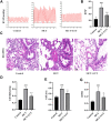
Luteolin effectively relieves MCT-induced PAH and pulmonary vascular remodeling. (A) Representative images of RVSP waveform (n = 6). (B) Statistical graph of RVSP (n = 6). (C) H. E. staining of pulmonary arterioles from control rats, MCT rats and MCT + LUT rats (400X) (n = 8) (D) Statistical graph of pulmonary vessel wall thickening (n = 8). (E) Statistical graph of pulmonary vessel wall area (n = 8). (F) Statistical graph of RVHI (n = 8). Scale: 50 μm, bars represent means ± SD, #control group vs MCT group, #p < 0.05, ##p < 0.01, *MCT group vs LUT group, *p < 0.05, **p < 0.01, ***p < 0.001, ****p < 0.0001. LUT, luteolin; MCT, monocrotaline; PAH, RVHI, right ventricular hypertrophy index; RVSP, right ventricular systolic pressure.
FIGURE 3
FIGURE 3
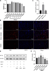
Luteolin inhibits the proliferation of rat PASMCs induced by PDGF-BB. PASMCs were cultured in 1% serum medium for 24 h, then treated with different concentrations of luteolin for 1 h before the stimulation with PDGF-BB (20 ng/ml). (A) Cell proliferation was examined using the Cell Counting kit eight test (n = 4); (B) and (C) DNA synthesis was examined using EdU, luteolin significantly inhibited the PDGF-BB induced DNA synthesis of PASMCs (n = 3). (D) and (E) the expression of protein level of PCNA was determined by western blot (n = 5). Scale: 50 μm, Bars represent means ± SD, #DMSO group vs PDGF + DMSO group, #p < 0.05, ##p < 0.01, *PDGF + DMSO group vs PDGF + LUT group, *p < 0.05, **p < 0.01, ***p < 0.001, ****p < 0.0001. DAPI, 4,6-diamino-2-phenylindole; EdU, 5-ethynyl-2′-deoxyuridine; LUT, luteolin; PASMCs, pulmonary arterial smooth muscle cells; PCNA, proliferating cell nuclear antigen; PDGF-BB, platelet-derived growth factor-BB.
FIGURE 4
FIGURE 4
Luteolin inhibits the migration of rat PASMCs induced by PDGF-BB. PASMCs were starved for 24 h in 1% serum medium, then treated with different concentrations of luteolin for 1 h before the stimulation with PDGF-BB (20 ng/ml). (A) Representative images from the wound healing assay of PASMCs (40X) (n = 3). (B) Statistical graph of wound healing experiment (n = 3). (C) and (D) the expression of protein level of MMP9 (n = 3). Bars represent means ± SD, #DMSO group vs PDGF + DMSO group, #p < 0.05, ##p < 0.01, *PDGF + DMSO group vs PDGF + LUT group, *p < 0.05, **p < 0.01, ***p < 0.001. MMP9, matrix metallopeptidase 9; LUT, luteolin; PASMCs, pulmonary arterial smooth muscle cells; PDGF-BB, platelet-derived growth factor-BB.
FIGURE 5
FIGURE 5
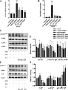
Luteolin inhibits the increase of mRNA and protein expression of key molecules in Hippo/YAP signaling pathway induced by PDGF-BB. PASMCs were cultured in 1% serum medium for 24 h, then treated with different concentrations of luteolin for 1 h before the stimulation with PDGF-BB (20 ng/ml). (A) mRNA level of LATS1 (n = 3). (B) mRNA level of YAP (n = 3). (C) and (D) the expression of protein level of LATS1 and phosphorylated LATS1 (n = 5). (E) and (F) the expression of protein level of YAP and phosphorylated YAP (n = 3). Bars represent means ± SD of n = 3–6, #DMSO group vs PDGF + DMSO group, #p < 0.05, ##p < 0.01, ###p < 0.0001, ###p < 0.0001, ####p < 0.00001, *PDGF + DMSO group vs PDGF + LUT group, *p < 0.05, **p < 0.01, ***p < 0.001, ****p < 0.0001, NS indicates no significance. LATS1, large tumor suppressor one; LUT, luteolin; PASMCs, pulmonary arterial smooth muscle cells; PDGF-BB, platelet-derived growth factor-BB; YAP, yes-association protein.
FIGURE 6
FIGURE 6
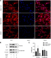
Luteolin decreases YAP nuclear localization induced by PDGF-BB. PASMCs were cultured in 1% serum medium for 24 h, then treated with luteolin (40 μM)for 1 h before the stimulation with PDGF-BB (20 ng/ml). The nuclear and cytoplasmic proteins are extracted separately and were measured by western blot analysis (A) Representative confocal microscopy images of immunofluorescence staining of YAP (red), luteolin decreased YAP nuclear localization induced by PDGF-BB (B) and (C) YAP levels in nuclear and cytoplasmic (n = 5). Scale: 20 μm, bars represent means ± SD, #DMSO group vs PDGF + DMSO group, #p < 0.05, *PDGF + DMSO group vs PDGF + LUT group, *p < 0.05. DAPI, 4,6-diamino-2-phenylindole; LUT, luteolin; PASMCs, pulmonary arterial smooth muscle cells; PDGF-BB, platelet-derived growth factor-BB; YAP, yes-association protein.
FIGURE 7
FIGURE 7
Luteolin inhibits PI3K/AKT signal pathway. PASMCs were cultured in 1% serum medium for 24 h, then treated with different concentrations of luteolin for 1 h before the stimulation with PDGF-BB (20 ng/ml). (A) and (B) The expression of protein level of PIK3CB was determined by western blot, luteolin reduced the expression of PIK3CB induced by PDGF-BB (n = 4). (C) and (D) The expression of protein level of total AKT and phosphorylated AKT and the ratio of p-AKT/AKT, luteolin significantly reduced the expression of phosphorylated AKT and the ratio of p-AKT/AKT induced by PDGF-BB (n = 5). Bars represent means ± SD of, #DMSO group vs PDGF + DMSO group, #p < 0.05, *PDGF + DMSO group vs PDGF + LUT group, *p < 0.05, NS indicates no significance. AKT, protein kinase B; LUT, luteolin; PASMCs, pulmonary arterial smooth muscle cells; PDGF-BB, platelet-derived growth factor-BB; PIK3CB, phosphatidylinositol 3-kinase catalytic beta polypeptide.

References

    1. Anson D. M., Wilcox R. M., Huseman E. D., Stump T. A., Paris R. L., Darkwah B. O., et al. (2018). Luteolin decreases epidermal growth factor receptor-mediated cell proliferation and induces apoptosis in glioblastoma cell lines. Basic Clin. Pharmacol. Toxicol. 123, 678–686. 10.1111/bcpt.13077 - DOI - PubMed
    1. Antoniu S. A. (2012). Targeting PDGF pathway in pulmonary arterial hypertension. Expert Opin. Ther. Targets 16, 1055–1063. 10.1517/14728222.2012.719500 - DOI - PubMed
    1. Barst R. J. (2005). PDGF signaling in pulmonary arterial hypertension. J. Clin. Invest. 115, 2691–2694. 10.1172/JCI26593 - DOI - PMC - PubMed
    1. Cao D., Zhu G.-Y., Lu Y., Yang A., Chen D., Huang H.-J., et al. (2020). Luteolin suppresses epithelial-mesenchymal transition and migration of triple-negative breast cancer cells by inhibiting YAP/TAZ activity. Biomed. Pharmacother. 129, 110462. 10.1016/j.biopha.2020.110462 - DOI - PubMed
    1. Chan C.-M., Chang H.-H., Wang V.-C., Huang C.-L., Hung C.-F. (2013). Inhibitory effects of resveratrol on PDGF-BB-induced retinal pigment epithelial cell migration via PDGFRβ, PI3K/akt and MAPK pathways. PLoS One 8, e56819, 10.1371/journal.pone.0056819 - DOI - PMC - PubMed

LinkOut - more resources