

Quantification and Monitoring of the Effect of Botulinum Toxin A on Paretic Calf Muscles of Children With Cerebral Palsy With MRI: A Preliminary Study
- PMID: 33935939
- PMCID: PMC8085320
- DOI: 10.3389/fneur.2021.630435
Quantification and Monitoring of the Effect of Botulinum Toxin A on Paretic Calf Muscles of Children With Cerebral Palsy With MRI: A Preliminary Study
Abstract
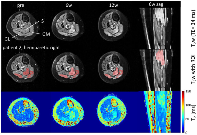
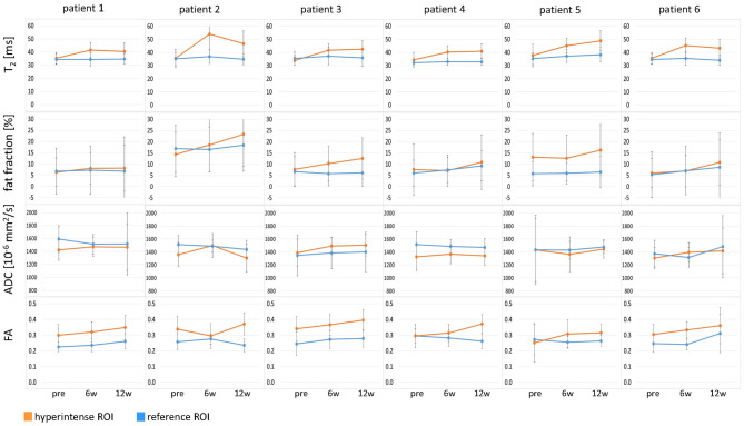
Background: Muscles from patients with cerebral palsy (CP) are often spastic and form contractures that limit the range of motion. Injections of botulinum toxin A (BTX) into the calf muscles are an important treatment for functional equinus; however, improvement in gait function is not always achieved. BTX is also used to test muscle weakening for risk evaluation of muscle lengthening surgery. Our aim was to assess the effect of BTX over time on calf muscle properties in pediatric CP patients with MRI. Material and Methods: Six toe-walking CP patients (mean age 11.6 years) with indication for lengthening surgery were prospectively enrolled and received BTX injections into the gastrocnemius and soleus muscles. MRI scans at 3T of the lower legs and clinical examinations were performed pre-BTX, 6 weeks (6w), and 12 weeks (12w) post-BTX. A fat-suppressed 2D multi-spin-echo sequence was used to acquire T2 maps and for segmentation. Fat fraction maps were calculated from 3D multi-echo Dixon images. Diffusion tensor imaging (DTI) with a 2D echo-planar imaging (EPI) sequence yielded maps of the mean apparent diffusion coefficient (ADC) and of the fractional anisotropy (FA). Hyperintense regions of interest (ROIs) on the T2-weighted (T2w) images at 6w were segmented in treated muscles. Mean values of T2, fat fraction, ADC, and FA were calculated in hyperintense ROIs and in reference ROIs in non-treated muscles. Results: Hyperintensity on T2w scans and increased T2 (group mean ± standard deviation: 35 ± 1 ms pre-BTX, 45 ± 2 ms at 6w, and 44 ± 2 ms at 12w) were observed in all patients at the injection sites. The T2 increase was spatially limited to parts of the injected muscles. FA increased (0.30 ± 0.03 pre-BTX, 0.34 ± 0.02 at 6w, and 0.36 ± 0.03 at 12w) while ADC did not change in hyperintense ROIs, indicating a BTX-induced increase in extracellular space and a simultaneous decrease of muscle fiber diameter. Fat fraction showed a trend for increase at 12w. Mean values in reference ROIs remained unchanged. Conclusion: MRI showed limited spatial distribution of the BTX-induced effects in pediatric CP patients. It could be a promising non-invasive tool for future studies to test BTX treatment protocols.
Keywords: MRI; T2; botulinum toxin A; calf muscles; cerebral palsy; diffusion; fat fraction; pediatric.
Copyright © 2021 Weidensteiner, Madoerin, Deligianni, Haas, Bieri, Akinci D'Antonoli, Bracht-Schweizer, Romkes, De Pieri, Santini, Rutz, Brunner and Garcia.
Conflict of interest statement
The authors declare that the research was conducted in the absence of any commercial or financial relationships that could be construed as a potential conflict of interest.
Figures


Similar articles
-
The Non-Affected Muscle Volume Compensates for the Partial Loss of Strength after Injection of Botulinum Toxin A.Toxins (Basel). 2023 Apr 3;15(4):267. doi: 10.3390/toxins15040267. Toxins (Basel). 2023. PMID: 37104205 Free PMC article.
-
Combination of Quantitative MRI Fat Fraction and Texture Analysis to Evaluate Spastic Muscles of Children With Cerebral Palsy.Front Neurol. 2021 Mar 22;12:633808. doi: 10.3389/fneur.2021.633808. eCollection 2021. Front Neurol. 2021. PMID: 33828520 Free PMC article.
-
Botulinum toxin a treatment in children with cerebral palsy: its effects on walking and energy expenditure.Am J Phys Med Rehabil. 2012 Jan;91(1):53-64. doi: 10.1097/PHM.0b013e31823caae1. Am J Phys Med Rehabil. 2012. PMID: 22157436
-
[Botulinum toxin treatment of hip adductor spasticity in multiple sclerosis].Wien Klin Wochenschr. 2001;113 Suppl 4:20-4. Wien Klin Wochenschr. 2001. PMID: 15506048 Review. German.
-
Botulinum toxin A treatment of the lower extremities in children with cerebral palsy.J Child Orthop. 2013 Nov;7(5):383-7. doi: 10.1007/s11832-013-0511-x. Epub 2013 Aug 28. J Child Orthop. 2013. PMID: 24432099 Free PMC article. Review.
Cited by
-
The Non-Affected Muscle Volume Compensates for the Partial Loss of Strength after Injection of Botulinum Toxin A.Toxins (Basel). 2023 Apr 3;15(4):267. doi: 10.3390/toxins15040267. Toxins (Basel). 2023. PMID: 37104205 Free PMC article.
-
Noninvasive analysis of overactive muscle structure and elasticity after botulinum toxin type A injection: a systematic review and meta-analysis.Eur J Phys Rehabil Med. 2024 Aug;60(4):567-580. doi: 10.23736/S1973-9087.24.08029-8. Epub 2024 Jul 3. Eur J Phys Rehabil Med. 2024. PMID: 38958691 Free PMC article.
-
Altered Muscle Contributions are Required to Support the Stance Limb During Voluntary Toe-Walking.Front Bioeng Biotechnol. 2022 Apr 11;10:810560. doi: 10.3389/fbioe.2022.810560. eCollection 2022. Front Bioeng Biotechnol. 2022. PMID: 35480978 Free PMC article.
-
Shear Wave Velocity to Evaluate the Effect of Botulinum Toxin on Post-Stroke Spasticity of the Lower Limb.Toxins (Basel). 2022 Dec 26;15(1):14. doi: 10.3390/toxins15010014. Toxins (Basel). 2022. PMID: 36668834 Free PMC article.
-
Triceps Surae Muscle Characteristics in Spastic Hemiparetic Stroke Survivors Treated with Botulinum Toxin Type A: Clinical Implications from Ultrasonographic Evaluation.Toxins (Basel). 2021 Dec 12;13(12):889. doi: 10.3390/toxins13120889. Toxins (Basel). 2021. PMID: 34941726 Free PMC article.
References
LinkOut - more resources
Full Text Sources
Other Literature Sources
Miscellaneous