Catheter-related giant right atrial thrombosis mimicking a myxoma: A case report
- PMID: 33936260
- PMCID: PMC8082616
- DOI: 10.3892/etm.2021.10035
Catheter-related giant right atrial thrombosis mimicking a myxoma: A case report
Abstract
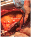
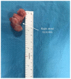
Despite the development of imagistic methods, the differential diagnosis of a right atrial mass may be difficult to be established, the most common pathologies which should be taken in consideration being represented by thrombus, tumors, prominent crista terminalis, or vegetation of infectious endocarditis. In this study, we present the case of a 63-year-old man with chronic kidney disease, in hemodialysis (HD) with a silicone central venous catheter (CVC) with the incidental transthoracic echocardiography (transthoracic echocardiogram, TTE) finding of a tumoral mass of 35x26 mm in the right atrium (RA), not related with the catheter, which was diagnosed as right atrial myxoma and underwent surgical excision. After reviewing the histopathology probe, the diagnosis of right atrial thrombus was confirmed. In conclusion, differentiating intracardiac right atrial masses (RAMs) could may prove challenging. In our patient, clinical presentation and the preoperative investigations could not differentiate the right atrial thrombus from a myxoma, and only the postoperative histopathology diagnosis was able to guide correct diagnosis.
Keywords: hemodialysis; myxoma; right atrial mass; right atriotomy; thrombus.
Copyright © 2020, Spandidos Publications.
Conflict of interest statement
The authors declare that they have no competing interests.
Figures


Similar articles
-
Diagnosis of the right atrial myxoma after treatment of COVID-19: A case report.Clin Case Rep. 2023 May 1;11(5):e7216. doi: 10.1002/ccr3.7216. eCollection 2023 May. Clin Case Rep. 2023. PMID: 37143454 Free PMC article.
-
Patient in chronic hemodialysis with right atrial mass: thrombus, fungal endocarditis or atrial myxoma?J Bras Nefrol. 2016 Dec;38(4):462-465. doi: 10.5935/0101-2800.20160073. J Bras Nefrol. 2016. PMID: 28001173 English, Portuguese.
-
An unusual presentation of prominent crista terminalis mimicking a right atrial mass: a case report.BMC Cardiovasc Disord. 2018 Nov 7;18(1):210. doi: 10.1186/s12872-018-0925-y. BMC Cardiovasc Disord. 2018. PMID: 30404609 Free PMC article.
-
Prominent crista terminalis mimicking a right atrial mass: a systematic literature review and meta-analysis.Acta Radiol. 2024 Jun;65(6):588-600. doi: 10.1177/02841851241242461. Epub 2024 Apr 15. Acta Radiol. 2024. PMID: 38619912
-
Over-the-Wire Retrieval of Infectious Hemodialysis Catheter-Related Right Atrial Thrombus Causing Recurrent Pleural Empyema and Sepsis: A Case-Based Review.J Clin Med. 2024 Nov 5;13(22):6630. doi: 10.3390/jcm13226630. J Clin Med. 2024. PMID: 39597776 Free PMC article. Review.
Cited by
-
Diagnosis of the right atrial myxoma after treatment of COVID-19: A case report.Clin Case Rep. 2023 May 1;11(5):e7216. doi: 10.1002/ccr3.7216. eCollection 2023 May. Clin Case Rep. 2023. PMID: 37143454 Free PMC article.
-
Management of catheter-related right atrial thrombus in hemodialysis: a systematic review.BMC Cardiovasc Disord. 2024 Nov 20;24(1):656. doi: 10.1186/s12872-024-04330-y. BMC Cardiovasc Disord. 2024. PMID: 39563254 Free PMC article.
-
Giant right atrial myxoma emerging from the suprahepatic inferior vena cava, extending to the right atrium; a case report and literature review.J Cardiothorac Surg. 2025 Aug 30;20(1):349. doi: 10.1186/s13019-025-03492-w. J Cardiothorac Surg. 2025. PMID: 40885975 Free PMC article. Review.
References
-
- McAllister HA Jr, Hall RJ, Cooley DA. Tumors of the heart and pericardium. Curr Probl Cardiol. 1999;24:57–116. - PubMed
-
- Cianciulli TF, Saccheri MC, Redruello HJ, Cosarinsky LA, Celano L, Trila CS, Parisi CE, Prezioso HA. Right atrial thrombus mimicking myxoma with pulmonary embolism in a patient with systemic lupus erythematosus and secondary antiphospholipid syndrome. Tex Heart Inst J. 2008;35:454–457. - PMC - PubMed
-
- Iliescu VA, Dorobantu LF, Stiru O, Iosifescu AG, Coman I, Marin S, Filipescu D. Second recurrence of cardiac myxoma 7 years after the initial operation. Chirurgia (Bucur) 2008;103:239–241. (In Romanian) - PubMed
LinkOut - more resources
Full Text Sources
Other Literature Sources
