

Cecal bascule with secondary gastric outlet obstruction in addition to a large bowel closed obstruction: A case report
- PMID: 33936351
- PMCID: PMC8079242
- DOI: 10.1016/j.radcr.2021.02.038
Cecal bascule with secondary gastric outlet obstruction in addition to a large bowel closed obstruction: A case report
Erratum in
-
Erratum regarding missing Declaration of Competing Interest statements in previously published articles.Radiol Case Rep. 2022 Sep 29;17(12):4933. doi: 10.1016/j.radcr.2022.08.054. eCollection 2022 Dec. Radiol Case Rep. 2022. PMID: 36311872 Free PMC article.
Abstract
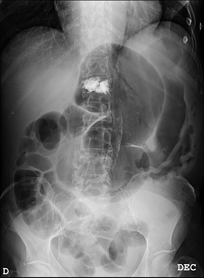
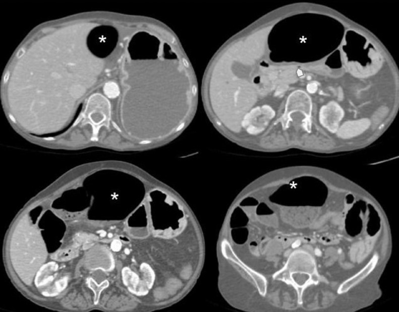
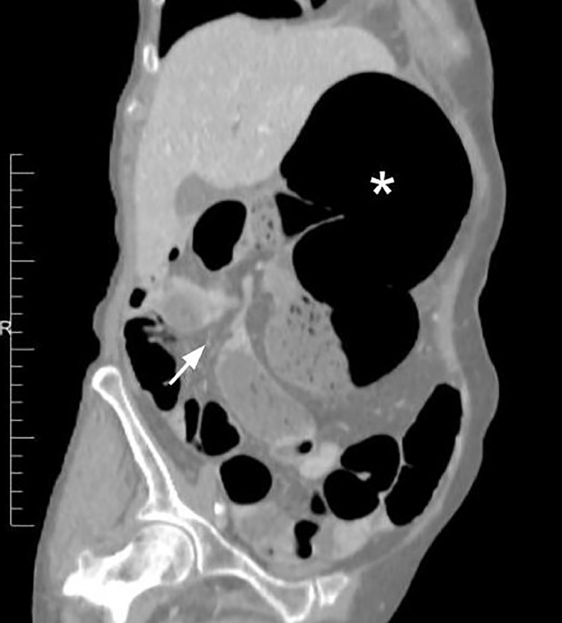
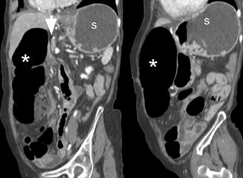
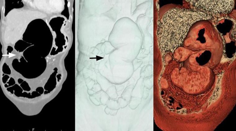
We present a case of cecal bascule in a 60-year-old woman with abdominal pain and vomiting. Imaging tests revealed a cecal bascule causing mechanic obstruction of the stomach. Besides a small bowel dilatation was not seen, the distended cecum was extrinsically obstructing the antrum and therefore, the gastric outlet. Cecal bascule is a form of cecal volvulus without the axial twisted component. The cecum folds anterior or anteromedially on itself and, because of a valve mechanism, becomes distended. The pressure increases and it begins to compromise arterial blood flow so life-threatening complications, such as bowel ischemia and infarction can occur. Prompt diagnosis is therefore crucial and plain radiographs and computed tomography are the preferred imaging techniques for this purpose.
Keywords: Acute abdomen; Diagnostic X-ray; Intestinal obstruction; Intestinal volvulus; Multidetector computed tomography.
© 2021 Published by Elsevier Inc. on behalf of University of Washington.
Figures
References
-
- Peterson CM, Anderson JS, Hara AK, Carenza JW, Menias CO. Volvulus of the gastrointestinal tract: appearances at multimodality imaging. Radiographics. 2009;29:1281–1293. - PubMed
-
- Moore CJ, Corl FM, Fishman EK. CT of cecal volvulus: unraveling the image. AJR Am J Roentgenol. 2001;177:95–98. - PubMed
-
- Frank AJ, Goffner LB, Fruauff AA, Losada RA. Cecal volvulus: the CT whirl sign. Abdom Imaging. 1993;18:288–289. - PubMed
Publication types
LinkOut - more resources
Full Text Sources
Other Literature Sources
