Pediatric follicular bronchiolitis with severe atelectasis: a case report
- PMID: 33936377
- PMCID: PMC8085817
Pediatric follicular bronchiolitis with severe atelectasis: a case report
Abstract
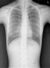
Follicular bronchiolitis is a rare pulmonary disorder characterized by the presence of multiple hyperplastic lymphoid follicles with a peribronchiolar distribution. An 11-year-old girl with total atelectasis of the right middle lobe (RML) and diffuse multiple small nodules at both lung bases presented to our hospital with frequent upper respiratory infections and pneumonia. The disease progressed during a 3-month period of macrolide therapy, and thoracoscopic biopsy with lobectomy of the atelectatic RML was performed. The histopathologic diagnosis was follicular bronchiolitis. The patient's pulmonary function improved dramatically after oral steroid treatment. It can be difficult to diagnose follicular bronchiolitis based solely on clinical, laboratory, and radiologic findings; the disorder must be confirmed histopathologically. A patient with longstanding irreversible atelectasis and resulting recurrent respiratory infection may require lobectomy for the diagnosis and treatment of follicular bronchiolitis.
Keywords: Follicular bronchiolitis; atelectasis; lobectomy; pediatrics.
IJCEP Copyright © 2021.
Conflict of interest statement
None.
Figures


Similar articles
-
Follicular bronchiolitis: A rare cause of persistent atelectasis in children.Respir Med Case Rep. 2013 Jul 10;10:7-9. doi: 10.1016/j.rmcr.2013.06.002. eCollection 2013. Respir Med Case Rep. 2013. PMID: 26029501 Free PMC article.
-
[Follicular bronchiolitis: report of 3 cases and literature review].Zhonghua Jie He He Hu Xi Za Zhi. 2017 Jun 12;40(6):457-462. doi: 10.3760/cma.j.issn.1001-0939.2017.06.012. Zhonghua Jie He He Hu Xi Za Zhi. 2017. PMID: 28592030 Review. Chinese.
-
HRCT findings of childhood follicular bronchiolitis.Pediatr Radiol. 2017 Dec;47(13):1759-1765. doi: 10.1007/s00247-017-3951-5. Epub 2017 Aug 26. Pediatr Radiol. 2017. PMID: 28844075
-
Follicular bronchiolitis: a rare disease in children.Turk Pediatri Ars. 2014 Dec 1;49(4):344-7. doi: 10.5152/tpa.2014.306. eCollection 2014 Dec. Turk Pediatri Ars. 2014. PMID: 26078687 Free PMC article.
-
[Bronchitis obliterans associated with bronchiolitis obliterans with organizing pneumonia in a child and literature review].Zhonghua Er Ke Za Zhi. 2016 Jul;54(7):523-6. doi: 10.3760/cma.j.issn.0578-1310.2016.07.010. Zhonghua Er Ke Za Zhi. 2016. PMID: 27412744 Review. Chinese.
References
-
- Ryu JH, Myers JL, Swensen SJ. Bronchiolar disorders. Am J Respir Crit Care Med. 2003;168:1277–1292. - PubMed
-
- Weinman JP, Manning DA, Liptzin DR, Krausert AJ, Browne LP. HRCT findings of childhood follicular bronchiolitis. Pediatr Radiol. 2017;47:1759–1765. - PubMed
-
- Yousem SA, Colby TV, Carrington CB. Follicular bronchitis/bronchiolitis. Hum Pathol. 1985;16:700–706. - PubMed
-
- Romero S, Barroso E, Gil J, Aranda I, Alonso S, Garcia-Pachon E. Follicular bronchiolitis: clinical and pathologic findings in six patients. Lung. 2003;181:309–319. - PubMed
Publication types
LinkOut - more resources
Full Text Sources
