

Case Reports
doi: 10.1002/ccr3.3665.
eCollection 2021 Apr.
Spontaneous healing in maxillary fungus ball: Beware of asymptomatic patients
Affiliations
- PMID: 33936605
- PMCID: PMC8077379
- DOI: 10.1002/ccr3.3665
Item in Clipboard
Case Reports
Spontaneous healing in maxillary fungus ball: Beware of asymptomatic patients
Clin Case Rep.
.
Abstract
Incidentally diagnosed, asymptomatic, paranasal fungus ball might occasionally undergo spontaneous healing. Accurate preoperative workup may avoid unnecessary surgical or anesthesiological procedures.
Keywords: computed tomography; dental disease; endoscopy; maxillary sinus; paranasal sinus; rhinosinusitis.
© 2020 The Authors. Clinical Case Reports published by John Wiley & Sons Ltd.
Conflict of interest statement
None declared.
Figures
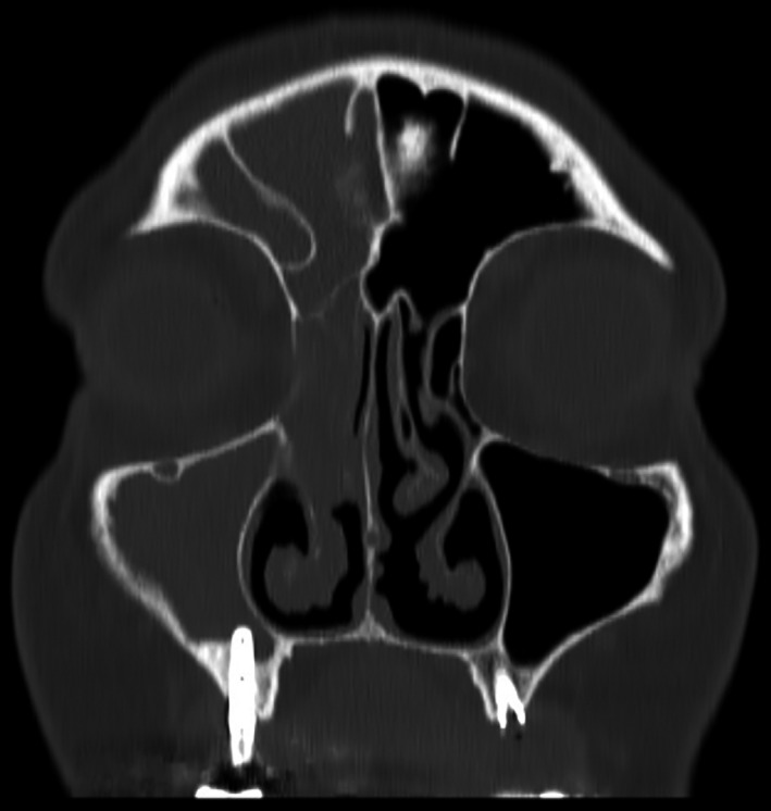
Coronal head CT scan image showing a right fronto‐ethmoid‐maxillary sinus opacification, with a dental implant protruding into the right maxillary sinus
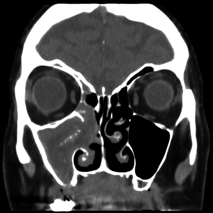
Coronal head CT scan image showing hyperdense floccular bodies floating amid the isodense opacity, suggesting the diagnosis of a paranasal sinus fungus ball
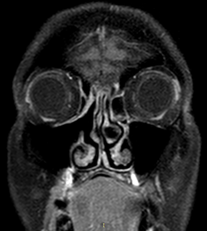
T1‐weighted coronal head MR image showing complete healing of the right maxillary sinus, with a wide antrostomy‐like aperture allowing for ventilation of the sinus
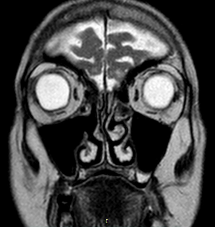
T2‐weighted coronal head MR image showing complete healing of the right maxillary sinus, with a wide antrostomy‐like aperture allowing for ventilation of the sinus
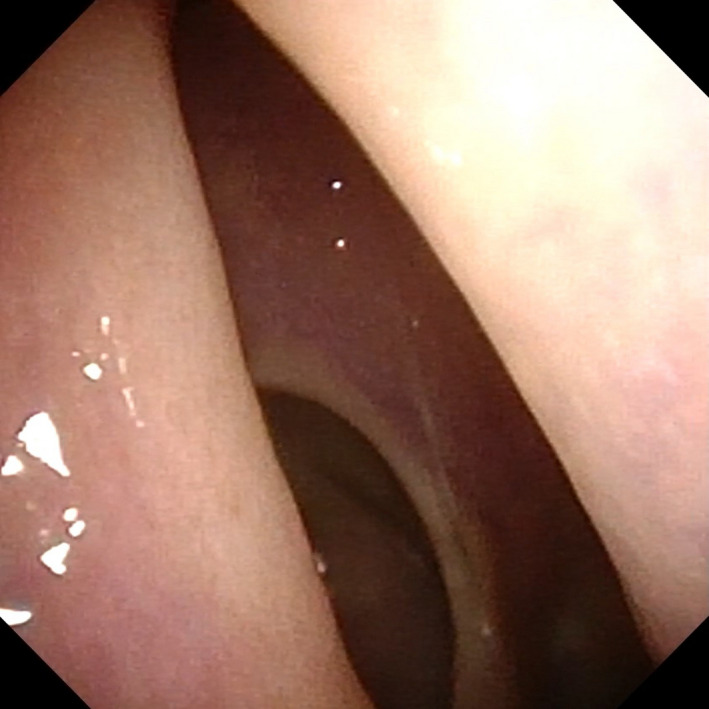
Flexible nasal endoscopy image of the right middle meatus showing a complete opening of the right maxillary sinus, without any residual sign of paranasal sinus fungus ball
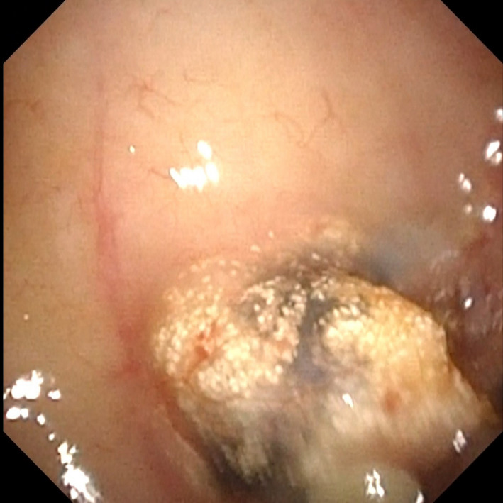
Flexible nasal endoscopy image of the right maxillary sinus floor, showing a small amount of granulomatous tissue in adherence with the protruding dental implant
References
-
- deShazo RD, O'Brien M, Chapin K, Soto‐Aguilar M, Gardner L, Swain R. A new classification and diagnostic criteria for invasive fungal sinusitis. Arch Otolaryngol Head Neck Surg. 1997;123:1181‐1188. - PubMed
-
- Kim DW, Kim YM, Min J‐Y, et al. Clinicopathologic characteristics of paranasal sinus fungus ball: retrospective, multicenter study in Korea. Eur Arch Otorhinolaryngol. 2020;277:761‐765. - PubMed
Publication types
LinkOut - more resources
Full Text Sources
