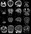
Cerebellar amelanotic melanoma can mimic cerebellar abscess in a pediatric case of neurocutaneous melanosis
- PMID: 33936629
- PMCID: PMC8077441
- DOI: 10.1002/ccr3.3926
Cerebellar amelanotic melanoma can mimic cerebellar abscess in a pediatric case of neurocutaneous melanosis
Abstract
Neurocutaneous melanosis (NCM) is a rare phakomatosis that may be associated with intracerebral masses. The differential diagnosis of intracerebral masses in NCM is often challenging and should include pigmented and nonpigmented lesions.
Keywords: adolescent medicine; neurology; paediatrics.
© 2021 The Authors. Clinical Case Reports published by John Wiley & Sons Ltd.
Conflict of interest statement
Authors declare no conflict of interest.
Figures


Similar articles
-
The pathologic features of neurocutaneous melanosis in a cynomolgus macaque.Vet Pathol. 2009 Jul;46(4):773-5. doi: 10.1354/vp.08-VP-0243-Q-BC. Epub 2009 Mar 9. Vet Pathol. 2009. PMID: 19276048
-
Imaging and Clinical Features of Neurocutaneous Melanosis in the Pediatric Population.Curr Med Imaging. 2021;17(12):1391-1402. doi: 10.2174/1573405617666210527091109. Curr Med Imaging. 2021. PMID: 34047260
-
Sudden change of a large congenital melanocytic nevus to neurocutaneous melanosis.J Craniofac Surg. 2006 Nov;17(6):1216-8. doi: 10.1097/01.scs.0000221514.34620.c0. J Craniofac Surg. 2006. PMID: 17119434
-
Neurosurgical management of patients with neurocutaneous melanosis: a systematic review.Neurosurg Focus. 2022 May;52(5):E8. doi: 10.3171/2022.2.FOCUS21791. Neurosurg Focus. 2022. PMID: 35535823
-
Neurocutaneous Melanosis in an Adult Patient with Intracranial Primary Malignant Melanoma: Case Report and Review of the Literature.World Neurosurg. 2018 Jun;114:76-83. doi: 10.1016/j.wneu.2018.02.007. Epub 2018 Mar 10. World Neurosurg. 2018. PMID: 29530698 Review.
Cited by
-
Ambient wing cistern: History, anatomy, imaging and approaches: An overview.World Neurosurg X. 2024 Sep 21;25:100395. doi: 10.1016/j.wnsx.2024.100395. eCollection 2025 Jan. World Neurosurg X. 2024. PMID: 39403178 Free PMC article. Review.
References
-
- Kadonaga JN, Frieden IJ. Neurocutaneous melanosis: Definition and review of the literature. J Am Acad Dermatol. 1991;24(5):747‐755. - PubMed
-
- van Heuzen EP, Kaiser MC, de Slegte RG, et al. Neurocutaneous melanosis. Pediatr Dermatol. 2002;23(3):747‐755.
-
- Abbo O, Dubedout S, Ballouhey Q, Maza A, Sevely A, Galinier P. Mélanose neurocutanée néonatale asymptomatique. Arch Pediatr. 2012;19(12):1319‐1321. - PubMed
-
- Byrd SE, Darling CF, Tomita T, Chou P, De Leon GA, Radkowski MA. MR imaging of symptomatic neurocutaneous melanosis in children. Pediatr Radiol. 1997;27(1):39‐44. - PubMed
-
- Shinno K, Nagahiro S, Uno M, et al. Neurocutaneous melanosis associated with malignant leptomeningeal melanoma in an adult:clinical significance of 5‐s‐cysteinyldopa in the cerebrospinal fluid. Neurol Med Chir (Tokyo). 2003;43(12):619‐625. - PubMed
Publication types
LinkOut - more resources
Full Text Sources
Other Literature Sources

