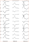
Electrocardiographic Criteria for Differentiating Left from Right Idiopathic Outflow Tract Ventricular Arrhythmias
- PMID: 33936738
- PMCID: PMC8076969
- DOI: 10.15420/aer.2020.10
Electrocardiographic Criteria for Differentiating Left from Right Idiopathic Outflow Tract Ventricular Arrhythmias
Abstract
Idiopathic ventricular arrhythmias are ventricular tachycardias or premature ventricular contractions presumably not related to myocardial scar or disorders of ion channels. Of the ventricular arrhythmias (VAs) without underlying structural heart disease, those arising from the ventricular outflow tracts (OTs) are the most common. The right ventricular outflow tract (RVOT) is the most common site of origin for OT-VAs, but these arrhythmias can, less frequently, originate from the left ventricular outflow tract (LVOT). OT-VAs are focal and have characteristic ECG features based on their anatomical origin. Radiofrequency catheter ablation (RFCA) is an effective and safe treatment strategy for OT-VAs. Prediction of the OT-VA origin according to ECG features is an essential part of the preprocedural planning for RFCA procedures. Several ECG criteria have been proposed for differentiating OT site of origin. Unfortunately, the ECG features of RVOT-VAs and LVOT-VAs are similar and could possibly lead to misdiagnosis. The authors review the ECG criteria used in clinical practice to differentiate RVOT-VAs from LVOT-VAs.
Keywords: Idiopathic ventricular arrhythmia; catheter ablation; electrocardiogram; ventricular outflow tract.
Copyright © 2021, Radcliffe Cardiology.
Conflict of interest statement
Disclosure: The authors have no conflicts of interest to declare.
Figures


References
-
- Yamada T,, Doppalapudi H,, McElderry HT, et al. Idiopathic ventricular arrhythmias originating from the papillary muscles in the left ventricle: prevalence, electrocardiographic and electrophysiological characteristics, and results of the radiofrequency catheter ablation. J Cardiovasc Electrophysiol. 2010;21:62–9. doi: 10.1111/j.1540-8167.2009.01594.x. - DOI - PubMed
Publication types
LinkOut - more resources
Full Text Sources

