. 2021 Apr 16:11:658608.
doi: 10.3389/fonc.2021.658608. eCollection 2021.

LCT-3d Induces Oxidative Stress-Mediated Apoptosis by Upregulating Death Receptor 5 in Gastric Cancer Cells

Affiliations

LCT-3d Induces Oxidative Stress-Mediated Apoptosis by Upregulating Death Receptor 5 in Gastric Cancer Cells

Menglin Wang et al. Front Oncol. .

Erratum in

Abstract

Gastric cancer is a global health problem. In this study, we investigate the role of a novel Indole derivative, named LCT-3d, in inhibiting the growth of gastric cancer cells by MTT assay. The Western blotting results showed that LCT-3d modulated the mitochondrial-related proteins and Cleaved-Caspases 3/9, to induce cell apoptosis. The up-regulation of Death receptor 5 (DR5) in MGC803 cells was observed with LCT-3d treatment. Knockdown of DR5 on MGC803 cells partially reversed the LCT-3d-induced mitochondrial apoptosis. The level of Reactive Oxygen Species (ROS) in MGC803 cells was increased with LCT-3d treatment and could be blocked with the pretreatment of the ROS inhibitor N-Acetylcysteine (NAC). The results demonstrate that the elevating ROS can up-regulate the expression of DR5, resulting in apoptosis via mitochondrial pathway. Although the nuclear factor erythroid-2 related factor 2 (Nrf2) pathway served an important role in protecting gastric cancer cells against the injury of ROS, it can't reverse LCT-3d-induced cell apoptosis. Taken together, our study showed that LCT-3d induced apoptosis via DR5-mediated mitochondrial apoptotic pathway in gastric cancer cells. LCT-3d could be a novel lead compound for development of anti-cancer activity in gastric cancer.

Keywords: DR5; LCT-3d; Nrf2; apoptosis; gastric cancer; reactive oxygen species.


Conflict of interest statement

The authors declare that the research was conducted in the absence of any commercial or financial relationships that could be construed as a potential conflict of interest. The reviewer JH declared a shared affiliation with one of the authors, G-YK, to the handling editor at time of review.

Figures

Figure 1
Figure 1
LCT-3d inhibited the cell proliferation of gastric cancer cells. (A) LCT-3d synthetic procedure. (B) Structure of LCT-3d. (C) Cytotoxic effect of LCT-3d on gastric cancer cells measured by MTT assay. Cells were treated with increasing concentration of LCT-3d for 48 h. *P < 0.05, **P < 0.01, significantly different compared with control.
Figure 2
Figure 2
LCT-3d triggered Caspase mediated apoptotic pathway in gastric cancer cells. (A) MGC803 cells and HGC27 were cells treated with various concentrations of LCT-3d for 48 h and apoptosis analyzed by flow cytometry. (B) Cells were treated as in (A) and the expression of Cleaved-Caspase and Cleaved PARP was analyzed by Western blotting. (C) MGC803 cells and HGC27 cells were pretreated with a pan-Caspase inhibitor, Z-VAD-FMK (100 μM) for 1 h, followed by incubation with LCT-3d (4 μM) for 48 h. Flow cytometric analysis on the effect of Z-VAD-FMK on LCT-3d-induced cells apoptosis.
Figure 3
Figure 3
LCT-3d-induced apoptosis was associated with mitochondrial pathway in gastric cancer cells. (A) MGC803 cells and HGC27 cells were treated with various concentrations of LCT-3d for 48 h and the membrane potential was measured by JC-1 dye retention using flow cytometry. (B) Cells were treated as in (A) and the expression of Bax, Bad, Bim, Bid, Bcl-xL, Bcl-2, and XIAP proteins was determined by Western blotting.
Figure 4
Figure 4
LCT-3d-induced apoptosis is associated with extrinsic pathway in gastric cancer cells. Both wild type and DR5−/− MGC803 cells were treated with 4 μM LCT-3d or indicated concentration. (A) Western blotting assay showed reduced expression of DR5 in a dose-dependent manner in MGC803 cells. (B) MGC803 cells and DR5−/− MGC803 cells were treated with 4 μM LCT-3d for 24 h and DR5 protein expressions in MGC803 cells determined by flow cytometry. (C) MTT assay showed the cell viability after treatment with 4 μM LCT-3d for 24 h. **P < 0.01, significantly different compared with control. (D) Flow cytometric analysis showed the ratios of apoptotic cells after treatment with 4 μM LCT-3d for 24 h. (E) Western blotting assay showed the expression of the apoptosis-related proteins after treatment with 4 μM LCT-3d for 24 h. (F) Flow cytometric analysis demonstrated the reduction of MMP (ΔΨ) after treatment with 4 μM LCT-3d for 24 h.
Figure 5
Figure 5
LCT-3d increased the level of ROS in gastric cancer cells. (A) MGC803 cells and HGC27 cells were treated with LCT-3d (4 μM) for indicated times and the level of ROS was detected by DCFH-DA with flow cytometry. (B) Western blotting assay showed Nrf2 nuclear-translocation and the changes of LCT-3d-induced protein expression at indicated time points. (C) MGC803 cells were treated with LCT-3d (4 μM) for 2 h. The treated and untreated samples are stained with Nrf2 antibody (Green) and DAPI (Blue) (magnification, 400×). The arrows indicate Nrf2 nuclear translocation.
Figure 6
Figure 6
LCT-3d-induced apoptosis is associated with the rising ROS in gastric cancer cells. (A) MGC803 cells and HGC27 cells were pretreated with NAC (5 mM) for 1 h, followed by incubation with LCT-3d (4 μM) for 48 h. The effect of NAC on LCT-3d-induced cell death was analyzed by MTT assay. **P < 0.01, significantly different compared with control. (B) Flow cytometric analysis demonstrated the effects of NAC (5 mM) on LCT-3d (4 μM)-induced cell apoptosis of the gastric cancer cells. (C) Western blotting assay showed the effect of NAC (5 mM) on LCT-3d (4 μM)-induced change of protein expression at 48 h. (D) Flow cytometric analysis showed the effect of NAC (5 mM) on LCT-3d (4 μM)-induced loss of MMP (ΔΨ) in gastric cancer cells. (E) MGC803 cells pretreated with ML385 for 1 h followed by the treatment with LCT-3d (4 μM) for an additional 48 h. The pretreatment with ML385 rarely reversed the cell death in MGC803 and HGC27 cells.
Figure 7
Figure 7
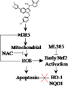
LCT-3d selectively kills gastric cancer cells but not normal cells via inducing ROS triggered apoptosis by activating death receptors and the mitochondria pathway. ROS scavenger NAC can block cell death, while Nrf2 inhibitor ML385 has no effect on LCT-3d-induced cell death. The promotion of ROS-induced apoptosis by LCT-3d depends on the up-regulation of DR5 but not the inactivation of Nrf2.

Similar articles

Cited by

References

    1. Thrift AP, El-Serag HB. Burden of Gastric Cancer. Clin Gastroenterol Hepatol (2020) 18:534–42. 10.1016/j.cgh.2019.07.045 - DOI - PMC - PubMed
    1. Cao J, Zhang X, Xu P, Wang H, Wang S, Zhang L, et al. Circular RNA circLMO7 acts as a microRNA-30a-3p sponge to promote gastric cancer progression via the WNT2/beta-catenin pathway. J Exp Clin Cancer Res (2021) 40:6. 10.1186/s13046-020-01791-9 - DOI - PMC - PubMed
    1. Shen X, Wang J, Yan X, Ren X, Wang F, Chen X, et al. Predictive value of GSTP1 Ile105Val polymorphism in clinical outcomes of chemotherapy in gastric and colorectal cancers: a systematic review and meta-analysis. Cancer Chemother Pharmacol (2016) 77:1285–302. 10.1007/s00280-016-3047-1 - DOI - PubMed
    1. Gao HL, Gupta P, Cui Q, Ashar YV, Wu ZX, Zeng L, et al. Sapitinib Reverses Anticancer Drug Resistance in Colon Cancer Cells Overexpressing the ABCB1 Transporter. Front Oncol (2020) 10:574861. 10.3389/fonc.2020.574861 - DOI - PMC - PubMed
    1. Lei ZN, Teng QX, Zhang W, Fan YF, Wang JQ, Cai CY, et al. Establishment and Characterization of a Topotecan Resistant Non-small Cell Lung Cancer NCI-H460/TPT10 Cell Line. Front Cell Dev Biol (2020) 8:607275. 10.3389/fcell.2020.607275 - DOI - PMC - PubMed

LinkOut - more resources