. 2021 Apr 15:8:674632.
doi: 10.3389/fmolb.2021.674632. eCollection 2021.

CRISPR/dCas9-Mediated Parkin Inhibition Impairs Mitophagy and Aggravates Apoptosis of Rat Nucleus Pulposus Cells Under Oxidative Stress

Affiliations

CRISPR/dCas9-Mediated Parkin Inhibition Impairs Mitophagy and Aggravates Apoptosis of Rat Nucleus Pulposus Cells Under Oxidative Stress

Tao Lan et al. Front Mol Biosci. .

Abstract

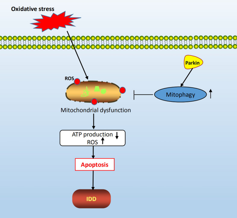
Objective: The aim of this study is to explore the role of Parkin in intervertebral disk degeneration (IDD) and its mitophagy regulation mechanism.

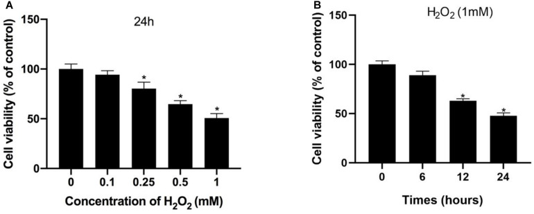
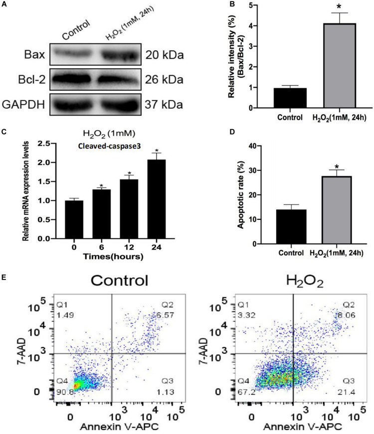
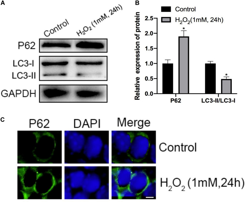
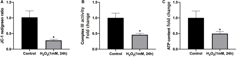
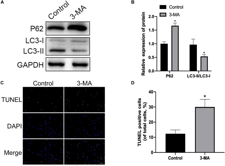
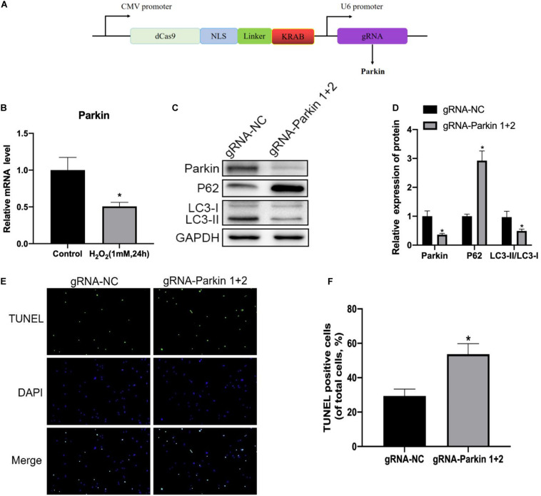
Study design and methods: Rat nucleus pulposus (NP) cells were stimulated with hydrogen peroxide (H2O2) to a mimic pathological condition. Apoptosis and mitophagy were assessed by Western blot, terminal deoxynucleotidyl transferase dUTP nick end labeling (TUNEL) assay, and immunofluorescence staining. The CRISPR-dCas9-KRAB system was used to silence the expression of Parkin.

Result: In this study, we found that Parkin was downregulated in rat NP cells under oxidative stress. In addition, treatment with H2O2 resulted in mitochondrial dysfunction, autophagy inhibition, and a significant increase in the rate of apoptosis of NP cells. Meanwhile, mitophagy inhibition enhanced H2O2-induced apoptosis. Furthermore, repression of Parkin significantly attenuated mitophagy and exacerbated apoptosis.

Conclusion: These results suggested that Parkin may play a protective role in alleviating the apoptosis of NP cells via mitophagy, and that targeting Parkin may provide a promising therapeutic strategy for the prevention of IDD.

Keywords: CRISPR/dCas9; IDD; Parkin; apoptosis; mitophagy.


Conflict of interest statement

The authors declare that the research was conducted in the absence of any commercial or financial relationships that could be construed as a potential conflict of interest.

Figures

FIGURE 1
FIGURE 1
Hydrogen peroxide inhibited cell viability in NP cells. (A,B) Effects of H2O2 on cell viability were detected using the Cell Counting Kit-8 (CCK-8). *p < 0.05 vs. control group.
FIGURE 2
FIGURE 2
Hydrogen peroxide promotes apoptosis in rat nucleus pulposus (NP) cells. (A,B) The protein levels of Bax and Bcl-2 in NP cells were measured by Western blotting. (C) Relative mRNA expression of cleaved caspase-3 by RT-qPCR. (D,E) Apoptosis of NP cells with or without H2O2 treatment was detected by flow cytometry. Data are represented as the mean ± SD. *p < 0.05 vs. control group.
FIGURE 3
FIGURE 3
Effects of H2O2 on mitophagy in NP cells. (A,B) The protein level of P62, LC3-I, and LC3-II in the NP cells was measured by Western blotting. (C) Immunofluorescence of the P62 protein in NP cells (Green signal represents P62, blue signal represents DAPI). *p < 0.05 vs. control group.
FIGURE 4
FIGURE 4
Mitochondrial dysfunction is involved in H2O2-induced apoptosis of rat NP. (A) Effect of H2O2 on mitochondrial membrane potential. (B) Summary data for the effect of H2O2 on the relative content of complex III. (C) Intracellular ATP levels in NP cells. *p < 0.05 vs. control group.
FIGURE 5
FIGURE 5
Mitophagy inhibition enhances H2O2-induced apoptosis in rat NP. (A,B) Western blot of P62, LC3-I, and LC3-II expression in NP cells after treatment with 3-MA. (C,D) Representative images of TUNEL staining and quantitative analysis showing NP cells apoptosis after treatment with 3-MA. *p < 0.05 vs. control group.
FIGURE 6
FIGURE 6
Inhibition of Parkin by the dCas9-KRAB system significantly attenuates H2O2-induced mitophagy. (A) Vector map of clustered regularly interspaced palindromic repeat epigenome editing vectors targeting the Parkin gene promotor region. (B) Expression level of Parkin in NP cells. (C,D) Western blot of Parkin, P62, LC3-I, and LC3-II expression in NP cells after Parkin inhibition. (E,F) Representative images of TUNEL staining and quantitative analysis showing NP cells apoptosis after Parkin inhibition. *p < 0.05 vs. control group.
FIGURE 7
FIGURE 7
Schematic diagram of mechanisms shows the protective role of mitophagy in rat NP cells under oxidative stress.

References

    1. Ansari M. Y., Khan N. M., Ahmad I., Haqqi T. M. (2018). Parkin clearance of dysfunctional mitochondria regulates ROS levels and increases survival of human chondrocytes. Osteoarthr. Cartil. 26 1087–1097. 10.1016/j.joca.2017.07.020 - DOI - PMC - PubMed
    1. Chen D., Xia D., Pan Z., Xu D., Zhou Y., Wu Y., et al. (2016). Metformin protects against apoptosis and senescence in nucleus pulposus cells and ameliorates disc degeneration in vivo. Cell Death Dis. 7:e2441. 10.1038/cddis.2016.334 - DOI - PMC - PubMed
    1. Chen Q., Chai Y. C., Mazumder S., Jiang C., Macklis R. M., Chisolm G. M., et al. (2003). The late increase in intracellular free radical oxygen species during apoptosis is associated with cytochrome c release, caspase activation, and mitochondrial dysfunction. Cell Death Differ. 10 323–334. - PMC - PubMed
    1. Chen S., Zhao L., Deng X., Shi D., Wu F., Liang H., et al. (2017). Mesenchymal stem cells protect nucleus pulposus cells from compression-induced apoptosis by inhibiting the mitochondrial pathway. Stem Cells Int. 2017:9843120. 10.1155/2017/9843120 - DOI - PMC - PubMed
    1. Chen Y., Lin J., Chen J., Huang C., Zhang Z., Wang J., et al. (2020). Mfn2 is involved in intervertebral disc degeneration through autophagy modulation. Osteoarthr. Carti. 28 363–374. 10.1016/j.joca.2019.12.009 - DOI - PubMed

LinkOut - more resources