Review
. 2021 Apr 15:8:614235.
doi: 10.3389/fcvm.2021.614235. eCollection 2021.

Novel Approaches in Cardiac Imaging for Non-invasive Assessment of Left Heart Myocardial Fibrosis

Affiliations
Review

Novel Approaches in Cardiac Imaging for Non-invasive Assessment of Left Heart Myocardial Fibrosis

Giulia Elena Mandoli et al. Front Cardiovasc Med. .

Abstract

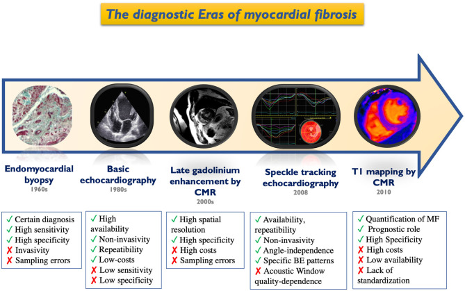
In the past, the identification of myocardial fibrosis was only possible through invasive histologic assessment. Although endomyocardial biopsy remains the gold standard, recent advances in cardiac imaging techniques have enabled non-invasive tissue characterization of the myocardium, which has also provided valuable insights into specific disease processes. The diagnostic accuracy, incremental yield and prognostic value of speckle tracking echocardiography, late gadolinium enhancement and parametric mapping modules by cardiac magnetic resonance and cardiac computed tomography have been validated against tissue samples and tested in broad patient populations, overall providing relevant clinical information to the cardiologist. This review describes the patterns of left ventricular and left atrial fibrosis, and their characterization by advanced echocardiography, cardiac magnetic resonance and cardiac computed tomography, allowing for clinical applications in sudden cardiac death and management of atrial fibrillation.

Keywords: cardiac magnetic resonance; echocardiography; fibrosis; myocardial strain; speckle tracking.


Conflict of interest statement

The authors declare that the research was conducted in the absence of any commercial or financial relationships that could be construed as a potential conflict of interest.

Figures

Figure 1
Figure 1
Historical timeline of cardiac imaging evolution for the detection of myocardial fibrosis. BE, bull-eye; CMR, cardiac magnetic resonance; MF, myocardial fibrosis.
Figure 2
Figure 2
Different patterns of left ventricular fibrosis in an acute myocardial infarction (left) and in acute myocarditis (right) as highlighted by impairment in segmental global longitudinal strain reduction at bull's eye and the corresponding region of late gadolinium enhancement.
Figure 3
Figure 3
Typical patterns of myocardial fibrosis in different cardiomyopathies by speckle tracking bull's eye representation (left) and by cardiac magnetic resonance late gadolinium enhancement.
Figure 4
Figure 4
Proposed diagnostic algorithm for the role of different imaging modalities e.g., cardiac magnetic resonance (CMR) in the diagnosis of disease causing left ventricular myocardial fibrosis with increased risk of sudden cardiac death. ARVD, arrhythmogenic right ventricular cardiomyopathy; CMR, cardiac magnetic resonance; HCM, hypertrophic cardiomyopathy; LGE, late gadolinium enhancement; LV, left ventricular.
Figure 5
Figure 5
Different features and applications of advanced imaging modalities for the evaluation of left atrial fibrosis: on the left, evaluation of left atrial deformation by speckle tracking echocardiography; on the right, left atrial 3D model rendering by cardiac magnetic resonance obtained tracing the left atrial wall and quantifying late gadolinium enhancement signal with different color coding (blue: normal tissue; green: fibrosis) [adapted from Siebermair et al. (97)].
Figure 6
Figure 6
Proposed algorithms integrating speckle tracking echocardiography (STE) and cardiac magnetic resonance (CMR) for the screening and management procedures of atrial fibrillation. AF, atrial fibrillation; AS, aortic stenosis; ECG, electrocardiogram; ECV, extracellular volume; HF, heart failure; LA, left atrial; MR, mitral regurgitation; OAC, oral anticoagulants.

References

    1. Moharram MA, Lamberts RR, Whalley G, Williams MJA, Coffey S. Myocardial tissue characterization using echocardiographic deformation imaging. Cardiovasc Ultrasound. (2019) 17:27. 10.1186/s12947-019-0176-9 - DOI - PMC - PubMed
    1. Bing R, Dweck MR. Myocardial fibrosis: why image, how to image and clinical implications. Heart. (2019) 105:1832–40. 10.1136/heartjnl-2019-315560 - DOI - PMC - PubMed
    1. Gyöngyösi M, Winkler J, Ramos I, Do QT, Firat H, McDonald K, et al. . Myocardial fibrosis: biomedical research from bench to bedside. Eur J Heart Fail. (2017) 19:177–91. 10.1002/ejhf.696 - DOI - PMC - PubMed
    1. Travers JG, Kamal FA, Robbins J, Yutzey KE, Blaxall BC. Cardiac fibrosis: the fibroblast awakens. Circ Res. (2016) 118:1021–40. 10.1161/CIRCRESAHA.115.306565 - DOI - PMC - PubMed
    1. Cooper LT, Baughman KL, Feldman AM, Frustaci A, Jessup M, Kuhl U, et al. . The role of endomyocardial biopsy in the management of cardiovascular disease: a scientific statement from the American Heart Association, the American College of Cardiology, and the European Society of Cardiology. Endorsed by the Heart Failure Society of America and the Heart Failure Association of the European Society of Cardiology. J Am Coll Cardiol. (2007) 50:1914–31. 10.1161/CIRCULATIONAHA.107.186093 - DOI - PubMed

LinkOut - more resources