. 2021 Apr 14;6(11):3892-3903.
doi: 10.1016/j.bioactmat.2021.03.034. eCollection 2021 Nov.

Self-curling electroconductive nerve dressing for enhancing peripheral nerve regeneration in diabetic rats

Affiliations

Self-curling electroconductive nerve dressing for enhancing peripheral nerve regeneration in diabetic rats

Can Liu et al. Bioact Mater. .

Abstract

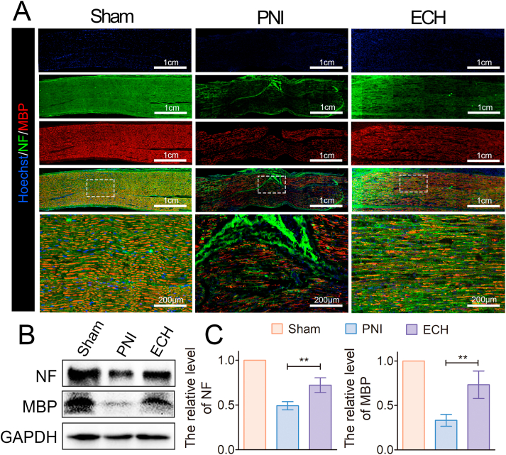
Conductive scaffolds have been shown to exert a therapeutic effect on patients suffering from peripheral nerve injuries (PNIs). However, conventional conductive conduits are made of rigid structures and have limited applications for impaired diabetic patients due to their mechanical mismatch with neural tissues and poor plasticity. We propose the development of biocompatible electroconductive hydrogels (ECHs) that are identical to a surgical dressing in this study. Based on excellent adhesive and self-healing properties, the thin film-like dressing can be easily attached to the injured nerve fibers, automatically warps a tubular structure without requiring any invasive techniques. The ECH offers an intimate and stable electrical bridge coupling with the electrogenic nerve tissues. The in vitro experiments indicated that the ECH promoted the migration and adhesion of the Schwann cells. Furthermore, the ECH facilitated axonal regeneration and remyelination in vitro and in vivo through the MEK/ERK pathway, thus preventing muscle denervation atrophy while retaining functional recovery. The results of this study are likely to facilitate the development of non-invasive treatment techniques for PNIs in diabetic patients utilizing electroconductive hydrogels.

Keywords: Axonal regeneration; Diabetic peripheral nerve injury; Electroconductive hydrogel; Nerve remyelination.


Conflict of interest statement

The authors declare that they have no known competing financial interests or personal relationships that could have appeared to influence the work reported in this paper.

Figures

Image 1
Graphical abstract
Fig. 1
Fig. 1
Characterization of ECHs. (A) Schematic of ECH formation and chemical structure. TA crosslinks with PPy chains by intermolecular electrostatic interactions and Fe3+ acts as an oxidant and ionic crosslinker. (B) SEM image of the ECH. (C) A photograph of dressing film plastered on the finger showed the adhesive properties of the ECH. (D) Illustrations and photographs showed self-curling ECH warped a size-matched tubular structure. (E–G) Electrical characterization, including (E) cyclic voltammograms, (F) Nyquist curves, and (G) Bode plots of ECH hydrogels showed excellent electrical performance. (H) The mechanistic properties of ECHs.
Fig. 2
Fig. 2
In vitro Schwann cells adhesion and migration on ECHs. (A) Live/dead assay of the SCs cultured on the hydrogel for one day. The green and red staining represent the live and dead cells, respectively. (B) The CCK-8 assay indicated that the cell viability exceeded 80% for the hydrogels after seeding (n = 5). (C) Cytoskeleton images demonstrated the adhesion of SCs cultured on each sample. (D) Wound-healing migration assay of the SCs on the hydrogels and control slide. (E) Quantitative analysis of the gap area at the denuded site (n = 3). (*p < 0.05, **p < 0.01, and ***p < 0.001). (For interpretation of the references to color in this figure legend, the reader is referred to the Web version of this article.)
Fig. 3
Fig. 3
Axonal outgrowth on the ECHs. (A) Immunocytochemistry of the PC-12 cells for the NF (green) and cell nuclei (blue) to analyze the axonal extension. (B) WB analysis to detect the NF and GAP43 expression values in PC-12 cells cultured in hydrogels and control conditions for 3 days. (C) Quantification of the integrated density for the protein bands (n = 3). (D–E) qRT-PCR data on the axonal gene expressions of the PC-12 cells. (F) Stained DRG for NF (green) and nuclei with DAPI (blue). (G–H) Analysis of the axonal lengths and distribution of the DRG for each group (n = 3). (*p < 0.05, **p < 0.01, and ***p < 0.001). (For interpretation of the references to color in this figure legend, the reader is referred to the Web version of this article.)
Fig. 4
Fig. 4
Functional analysis and morphological evaluation of the regenerated nerves. (A) Hydrogel implantation in a diabetic rat with sciatic nerve defect. (B) Structural repair indicated by H&E staining of the sciatic nerve on the 28th day after the injury. (C) The equation for calculating the SFI and measurement parameters of the footprints. (D) SFI scores of different groups 14, 21, and 28 days after the operation (n = 5). (E) Representative analysis of the footprints. (F) Illustration of the CMAP testing protocol. (G) Quantification of the CMAP amplitudes measured in each group (n = 3). (H) Representative CMAP recordings on the injured side of the sciatic nerve (*p < 0.05, **p < 0.01, and ***p < 0.001).
Fig. 5
Fig. 5
Promotion of axonal regeneration by ECH treatment in diabetic PNI models. (A) Immunocytochemistry of the NF (green) and MBP (red) proteins in the sciatic nerve tissues on the 28th day after the injury. (B) WB analysis of the NF and MBP protein bands in the sham, PNI, and hydrogel groups. (C) Quantitative measurement of the protein band intensity of the NF and MBP proteins (n = 3) (*p < 0.05, **p < 0.01, and ***p < 0.001). (For interpretation of the references to color in this figure legend, the reader is referred to the Web version of this article.)
Fig. 6
Fig. 6
Axonal remyelination of the regenerated axons after ECH treatment. (A) Immunocytochemistry of the S-100β (red) proteins in the regenerated tissues on the 28th day after the injury. (B) Expression levels of the S-100β protein in each group. (C) Quantitative analysis of western blot (n = 3) (D) Expression levels of the MEK/ERK pathway proteins. (E–F) Quantitative analysis of the protein expression, normalized to the sham group (n = 3). (*p < 0.05, **p < 0.01, and ***p < 0.001). (For interpretation of the references to color in this figure legend, the reader is referred to the Web version of this article.)
Fig. 7
Fig. 7
Attenuation of gastrocnemius muscle atrophy after ECH treatment. (A) Gross images of the isolated gastrocnemius muscles in each group. (B) The cross-sectional view of the ipsilateral muscles highlighted by Masson's trichrome stain. (C–E) Statistical analysis of the (C) wet weight ratio of the gastrocnemius muscle, (D) percentage of muscle fiber, and (E) percentage of collagen deposits area (n = 3) (*p < 0.05, **p < 0.01, and ***p < 0.001).
Fig. 8
Fig. 8
Morphological evaluation of the myelination of the regenerated axons. (A) Toluidine blue staining indicating remyelination of the regenerated nerve (B) Representative TEM images of the regenerated sciatic nerve. (C–F) Histomorphometric analysis of the remyelination of the regenerated axons through four parameters, namely (C) myelinated axon diameter, (D) myelinated axon density, (E) myelin sheath thickness, and (F) G-Ratio (n = 3) (*p < 0.05, **p < 0.01, and ***p < 0.001). (For interpretation of the references to color in this figure legend, the reader is referred to the Web version of this article.)

References

    1. Whiting D.R., Guariguata L., Weil C., Shaw J. IDF diabetes atlas: global estimates of the prevalence of diabetes for 2011 and 2030. Diabetes Res. Clin. Pract. 2011;94(3):311–321. - PubMed
    1. Feldman E.L., Nave K.-A., Jensen T.S., Bennett D.L.H. New horizons in diabetic neuropathy: mechanisms, bioenergetics, and pain. Neuron. 2017;93(6):1296–1313. - PMC - PubMed
    1. Callaghan B.C., Cheng H.T., Stables C.L., Smith A.L., Feldman E.L. Diabetic neuropathy: clinical manifestations and current treatments. Lancet Neurol. 2012;11(6):521–534. - PMC - PubMed
    1. Zenker J., Ziegler D., Chrast R. Novel pathogenic pathways in diabetic neuropathy. Trends Neurosci. 2013;36(8):439–449. - PubMed
    1. Li R., Li Y., Wu Y., Zhao Y., Chen H., Yuan Y., Xu K., Zhang H., Lu Y., Wang J., Li X., Jia X., Xiao J. Heparin-poloxamer thermosensitive hydrogel loaded with bFGF and NGF enhances peripheral nerve regeneration in diabetic rats. Biomaterials. 2018;168:24–37. - PMC - PubMed

LinkOut - more resources