. 2021 Sep;23(9):e13350.
doi: 10.1111/cmi.13350. Epub 2021 Jun 9.

Pathogenesis of Borrelia burgdorferi and Babesia microti in TLR4-Competent and TLR4-dysfunctional C3H mice

Affiliations

Pathogenesis of Borrelia burgdorferi and Babesia microti in TLR4-Competent and TLR4-dysfunctional C3H mice

Lavoisier Akoolo et al. Cell Microbiol. 2021 Sep.

Abstract

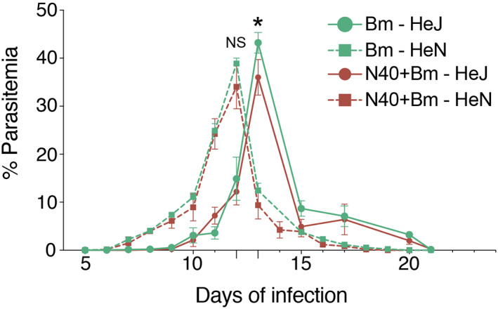
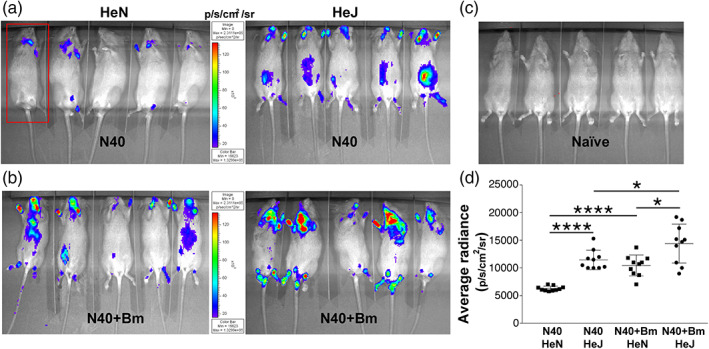
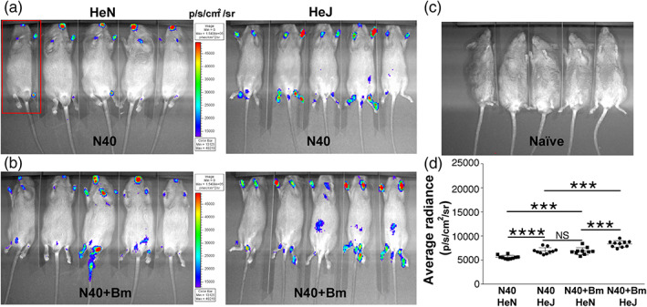
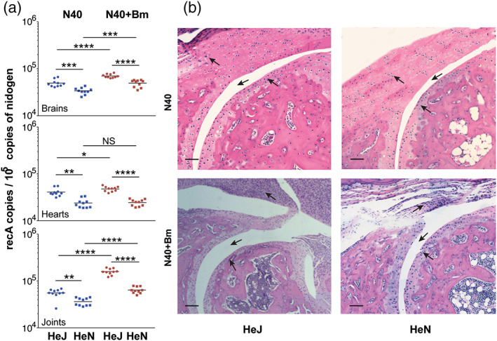
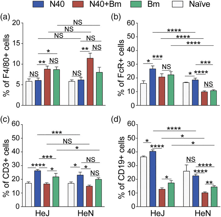
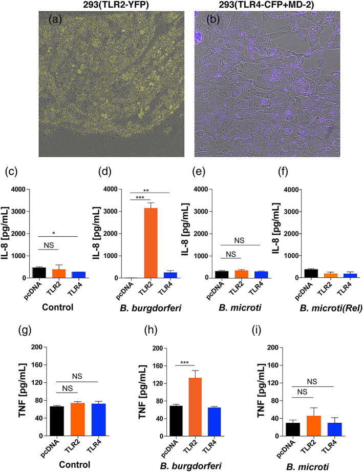
Toll-like receptors (TLRs) are a class of membrane-spanning proteins of host cells. TLR2 and TLR4 are displayed on the surface of macrophages, neutrophils and dendritic cells and recognise structurally conserved microbial signatures defined as Pathogen associated molecular patterns (PAMPs). C3H mice are susceptible to tick-borne pathogens; Lyme disease causing Borrelia burgdorferi that manifests arthritis and carditis and Apicomplexan protozoan, Babesia microti (Bm) that causes significant parasitemia associated with erythrocytopenia and haemoglobinuria. B. burgdorferi lacks typical TLR4 ligand lipopolysaccharides (LPS) and Bm TLR ligand(s) remain unknown. Only Borrelia lipoproteins that signal through TLR2 are established as PAMPs of these pathogens for TLR2/TLR4. Infection of C3H mice with each pathogen individually resulted in increase in the percentage of splenic B, T and FcR+ cells while their co-infection significantly diminished levels of these cells and caused increased B. burgdorferi burden in the specific organs. The most pronounced inflammatory arthritis was observed in co-infected C3H/HeJ mice. Parasitemia levels and kinetics of resolution of Bm in both mice strains were not significantly different. Transfected HEK293 cells showed pronounced signalling by B. burgdorferi through TLR2 and to some extent by TLR4 while Bm and infected erythrocytes did not show any response confirming our results in mice.

Keywords: Babesia microti; Babesiosis; Borrelia burgdorferi; Lyme disease; co-infection; inflammation; toll like receptor 4.


Conflict of interest statement

The authors declare that the research was conducted in the absence of any commercial or financial relationships that could be construed as a potential conflict of interest.

Figures

FIGURE 1
FIGURE 1
TLR4 deficiency prolongs survival of B microti in mouse blood. Parasitemia determined in 10 C3H/HeJ and C3H/HeN mice infected with Bm (104 infected erythrocytes injected i.p.) alone, or together with B. burgdorferi (103 spirochetes injected s.c. per mouse) by microscopic examination of Giemsa‐stained thin blood smears at regular intervals. Each point represents average parasitemia in each group of mice (mean ± SD) with peak parasitemia significantly higher (p < .05) in Bm infected than in respective co‐infected mouse strain but not significantly different between C3H/HeJ and C3H/HeN mice due to each infection (NS). Onset of parasitemia in Bm infected and co‐infected C3H/HeN appeared earlier than in C3H/HeJ mice (7th vs. 10th day post‐infection) but it cleared slightly faster (on 19th and 18th day of infection, respectively) than that in C3H/HeJ mice in which parasitemia was detectable in both Bm infected and co‐infected mice until 20th day post‐infection
FIGURE 2
FIGURE 2
Colonisation of organs by B. burgdorferi in C3H/HeJ and C3H/HeN mice infected with N40 alone and co‐infected with Bm at 2 weeks of infection. (a) Representative real‐time images of five C3H/HeJ (HeJ) and C3H/HeN (HeN) mice infected with bioluminescent N40 strain using IVIS‐200 displaying bioluminescence as a semi‐quantitative indicator of B. burgdorferi colonisation in different tissues of mice. Bioluminescence radiance in the whole body of each mouse (as marked in one mouse by a red rectangle) was also measured. (b) Overall, light emission in five mice each, representing colonisation of organs and spirochetal burden in co‐infected C3H/HeJ and C3H/HeN mice at 2 weeks post‐infection. (c) A set of five uninfected mice was also imaged at the same time with average bioluminescence radiance measured to be (483.23 p/s/cm2/sr). (d) Average bioluminescence radiance from five uninfected mice was deducted from the radiance obtained for each infected mouse (as marked in 2a and 2b) and net radiance values data for 10 mice for each infection is presented. Horizontal lines indicate the net mean radiance in each group of infected mice. Statistical analysis was conducted using a two‐tailed unpaired student t tests for unequal variance to determine significant difference between the paired groups (*p < .05, ****p < .0001)
FIGURE 3
FIGURE 3
Colonisation of mice organs by B. burgdorferi as detected by light emission due to the presence of live bioluminescent N40 strain at 3 weeks of infection. (a) Representative real‐time images of five C3H/HeJ (HeJ) and C3H/HeN (HeN) mice infected with bioluminescent N40 strain using IVIS‐200 displaying bioluminescence in the head and joints regions. Bioluminescence radiance in the whole body of each mouse (as marked in one mouse by a red rectangle) was also measured. (b) Bioluminescence detection in co‐infected C3H/HeJ and C3H/HeN mice. (c) Live imaging of a set of five uninfected mice at the same time point. (d) Average bioluminescence radiance from five uninfected mice was deducted from the radiance obtained for each infected mouse and net radiance data for each mouse is shown. Horizontal lines indicate the comparison of net mean radiance in each group of infected mice. Difference between average bioluminescence radiance in N40‐infected C3H/HeJ and co‐infected C3H/HeN mice was not statistically significant (NS). Statistical analysis was conducted using a two‐tailed unpaired student t tests for unequal variance to determine significant difference between the paired groups (***p < .001, ****p < .0001)
FIGURE 4
FIGURE 4
Borrelia burgdorferi strain N40 colonisation levels determined by qPCR and resulting pathology in C3H/HeJ and C3H/HeN mice. (a) Burden of B. burgdorferi in tissues as detected by duplex qPCR assay from 10 mice for each group using recA primers and molecular beacon probes with mouse nidogen amplicon copy number used for normalisation of spirochete quantities. Horizontal lines represent the mean B. burgdorferi recA copy number per 105 copies of nidogen genes. Statistical analysis was conducted using a two‐tailed unpaired student t tests for unequal variance to determine significant difference between the paired groups (NS‐Not significant, *p < .05, **p < .01, ***p < .001, ****p < .0001). (b) Severe arthritis in tibiotarsal joint manifested by synovial hyperplasia and erosion of cartilage, higher lymphocytic infiltration and change in synovial space (arrows) were observed in co‐infected mice as compared to B. burgdorferi‐infected mice of both strains. Bar represents a size of 100 μm
FIGURE 5
FIGURE 5
Modulation of splenic immune responses in mice by infection with B microti and N40 separately, or simultaneously. (a) Single cell suspension of mice spleen infected with N40, Bm and N40 + Bm were prepared and stained with fluorophore‐conjugated antibodies against F4/80+ (macrophages), (b) FcR+ phagocytic cells, (c) CD3+ (T cells), and (d) CD19+ (B cells), followed by FACS analysis. Higher percentages of B, FcR+ phagocytic and T cells were observed in C3H/HeJ mice. Proportions of various immune cells from spleens of mice from each experimental group analysed by FACS are expressed as mean ± SD. Statistical analysis was conducted using a two‐tailed unpaired student t tests for unequal variance to determine significant difference between the paired groups (NS‐Not Significant, *p < .05, **p < .01, ***p < .001, ****p < .0001)
FIGURE 6
FIGURE 6
Response of TLR2/TLR4 transfected HEK293 cells to B. burgdorferi N40 strain and Bm‐infected mouse blood. (a and b) Yellow fluorescence and Cyan fluorescence associated with TLR2‐YFP and TLR4‐CFP + MD‐2 transfected HEK293 cells confirmed expression of respective TLRs. (c and d) As compared to control (6c, medium only) treatment, exposure of transfected HEK293 cells to 2 × 107 B. burgdorferi for ~16 hr resulted in high stimulation of TLR2 and moderate but significant level of stimulation of TLR4 to produce pro‐inflammatory IL‐8 cytokine (6d). (e and f) Neither treatment with 1 × 106 RBCs of Bm infected mouse blood (~3.1 × 105 iRBCs) nor released (Rel) Bm parasites suspended in 20 μg/ml Polymyxin B containing treatment medium stimulated TLR2 or TLR4 to produce pro‐inflammatory IL‐8 cytokine. (g–i) Significant TNF‐α (TNF) stimulation was only observed by B. burgdorferi N40 strain treatment of HEK293 cells transfected with TLR2. Again, Bm did not shown stimulation neither of TLR2 nor TLR4 expressed in HEK293 cells. Statistical analysis was conducted using a two‐tailed unpaired student t tests for unequal variance to determine significant difference between the paired groups (NS‐Not significant, *p < .05, **p < .01, ****p < .0001)

References

    1. Abdollahi‐Roodsaz, S., Koenders, M. I., Walgreen, B., Bolscher, J., Helsen, M. M., van den Bersselaar, L. A., … van den Berg, W. B. (2013). Toll‐like receptor 2 controls acute immune complex‐driven arthritis in mice by regulating the inhibitory Fcgamma receptor IIB. Arthritis and Rheumatism, 65(10), 2583–2593. 10.1002/art.38087 - DOI - PubMed
    1. Aliota, M. T., Dupuis, A. P., 2nd, Wilczek, M. P., Peters, R. J., Ostfeld, R. S., & Kramer, L. D. (2014). The prevalence of zoonotic tick‐borne pathogens in Ixodes scapularis collected in the Hudson Valley, New York state. Vector Borne and Zoonotic Diseases, 14(4), 245–250. 10.1089/vbz.2013.1475 - DOI - PMC - PubMed
    1. Armstrong, A. L., Barthold, S. W., Persing, D. H., & Beck, D. S. (1992). Carditis in Lyme disease susceptible and resistant strains of laboratory mice infected with Borrelia burgdorferi . The American Journal of Tropical Medicine and Hygiene, 47(2), 249–258. - PubMed
    1. Anderson, J. F., Mintz, E. D., Gadbaw, & J. J. , Magnarelli, L. A. (1991). Babesia microti, human babesiosis, and Borrelia burgdorferi in Connecticut. Journal of Clinical Microbiology, 29(12), 2779–2783. 10.1128/jcm.29.12.2779-2783.1991. - DOI - PMC - PubMed
    1. Belongia, E. A. (2002). Epidemiology and Impact of Coinfections Acquired from Ixodes Ticks. Vector‐Borne and Zoonotic Diseases, 2(4), 265–273. 10.1089/153036602321653851. - DOI - PubMed

Publication types