

Development of an L-band resonator optimized for fast scan EPR imaging of the mouse head
- PMID: 33938574
- PMCID: PMC8295191
- DOI: 10.1002/mrm.28821
Development of an L-band resonator optimized for fast scan EPR imaging of the mouse head
Abstract
Purpose: To develop a novel resonator for high-quality fast scan electron paramagnetic resonance (EPR) and EPR/NMR co-imaging of the head and brain of mice at 1.25 GHz.
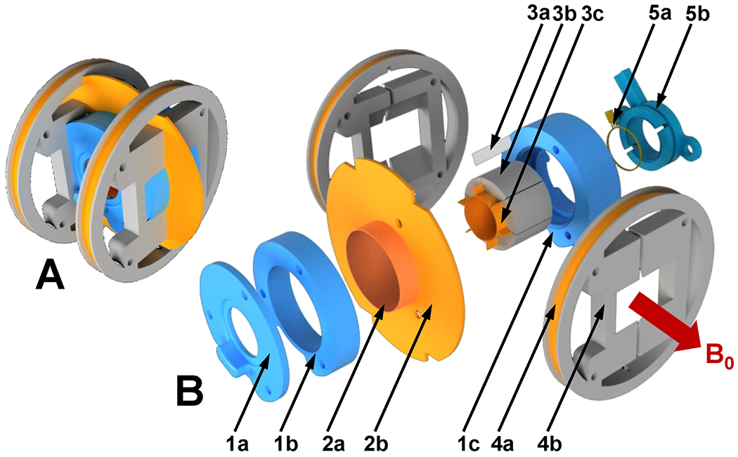
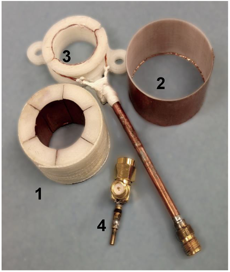
Methods: Resonator dimensions were scaled to fit the mouse head with maximum filling factor. A single-loop 6-gap resonator of 20 mm diameter and 20 mm length was constructed. High resonator stability was achieved utilizing a fixed position double coupling loop. Symmetrical mutually inverted connections rendered it insensitive to field modulation and fast scan. Coupling adjustment was provided by a parallel-connected variable capacitor located at the feeding line at λ/4 distance. To minimize radiation loss, the shield around the resonator was supplemented with a planar conductive disc that focuses return magnetic flux.
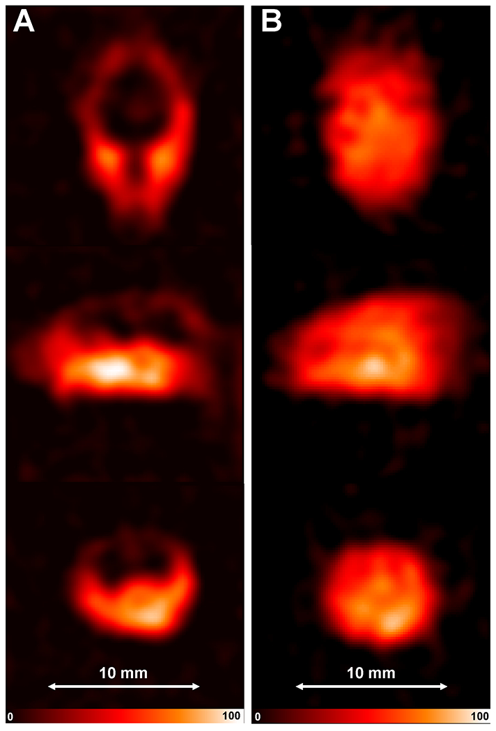
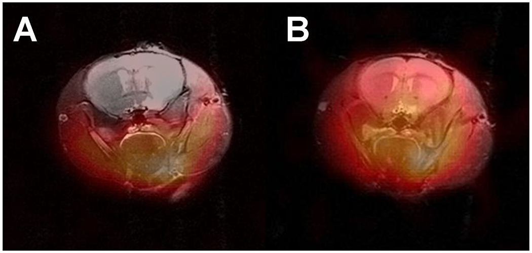
Results: Coupling of the resonator loaded with the mouse head was efficient and easy. This resonator enabled high-quality in vivo 3D EPR imaging of the mouse head following intravenous infusion of nitroxide probes. With this resonator and rapid scan EPR system, 4 ms scans were acquired in forward and reverse directions so that images with 2-scan 3,136 projections were acquired in 25 s. Head images were achieved with resolutions of 0.4 mm, enabling visualization of probe localization and uptake across the blood-brain barrier.
Conclusions: This resonator design provides good sensitivity, high stability, and B1 field homogeneity for in vivo fast scan EPR of the mouse head and brain, enabling faster measurements and higher resolution imaging of probe uptake, localization, and metabolism than previously possible.
Keywords: electron paramagnetic resonance imaging; instrument development; microwave resonator; mouse head imaging; paramagnetic probes.
© 2021 International Society for Magnetic Resonance in Medicine.
Figures


Similar articles
-
Single loop multi-gap resonator for whole body EPR imaging of mice at 1.2 GHz.J Magn Reson. 2007 Sep;188(1):68-73. doi: 10.1016/j.jmr.2007.05.021. Epub 2007 Jun 9. J Magn Reson. 2007. PMID: 17625940 Free PMC article.
-
Three-dimensional whole-body imaging of the bioreduction and clearance of nitroxide probes in the thoracic and abdominal regions of mice using a compact and mobile electron paramagnetic resonance imager.Magn Reson Med. 2025 Jul;94(1):424-435. doi: 10.1002/mrm.30432. Epub 2025 Jan 20. Magn Reson Med. 2025. PMID: 39831441
-
Development of a fast-scan EPR imaging system for highly accelerated free radical imaging.Magn Reson Med. 2019 Aug;82(2):842-853. doi: 10.1002/mrm.27759. Epub 2019 Apr 25. Magn Reson Med. 2019. PMID: 31020713 Free PMC article.
-
Rapid-scan EPR imaging.J Magn Reson. 2017 Jul;280:140-148. doi: 10.1016/j.jmr.2017.02.013. J Magn Reson. 2017. PMID: 28579099 Free PMC article. Review.
-
Measurement of oxygen concentrations in the intact beating heart using electron paramagnetic resonance spectroscopy: a technique for measuring oxygen concentrations in situ.J Bioenerg Biomembr. 1991 Dec;23(6):855-71. doi: 10.1007/BF00786005. J Bioenerg Biomembr. 1991. PMID: 1663949 Review.
Cited by
-
Rapid scan EPR: Automated digital resonator control for low-latency data acquisition.J Magn Reson. 2022 Dec;345:107308. doi: 10.1016/j.jmr.2022.107308. Epub 2022 Oct 21. J Magn Reson. 2022. PMID: 36356489 Free PMC article.
-
Electron Spin Resonance Probe Incorporation into Bioinks Permits Longitudinal Oxygen Imaging of Bioprinted Constructs.Mol Imaging Biol. 2024 Jun;26(3):511-524. doi: 10.1007/s11307-023-01871-0. Epub 2023 Dec 1. Mol Imaging Biol. 2024. PMID: 38038860 Free PMC article.
-
High fidelity triangular sweep of the magnetic field for millisecond scan EPR imaging.J Magn Reson. 2021 Aug;329:107024. doi: 10.1016/j.jmr.2021.107024. Epub 2021 Jun 9. J Magn Reson. 2021. PMID: 34198184 Free PMC article.
References
-
- Fujii HG, Emoto MC, Sato-Akaba H. Brain Redox Imaging Using In Vivo Electron Paramagnetic Resonance Imaging and Nitroxide Imaging Probes. Magnetochemistry 2019;5(11).
-
- Matsumura A, Emoto MC, Suzuki S, Iwahara N, Hisahara S, Kawamata J, Suzuki H, Yamauchi A, Sato-Akaba H, Fujii HG, Shimohama S. Evaluation of oxidative stress in the brain of a transgenic mouse model of Alzheimer disease by in vivo electron paramagnetic resonance imaging. Free Radical Biology & Medicine 2015;85:165–173. - PubMed
-
- Emoto MC, Sato-Akaba H, Matsuoka Y, Yamada KI, Fujii HG. Non-invasive mapping of glutathione levels in mouse brains by in vivo electron paramagnetic resonance (EPR) imaging: Applied to a kindling mouse model. Neuroscience Letters 2019;690:6–10. - PubMed
Publication types
MeSH terms
Grants and funding
LinkOut - more resources
Full Text Sources
Other Literature Sources
Medical
Miscellaneous
