

A comparison of breast and lung doses from chest CT scans using organ-based tube current modulation (OBTCM) vs. Automatic tube current modulation (ATCM)
- PMID: 33939253
- PMCID: PMC8130227
- DOI: 10.1002/acm2.13198
A comparison of breast and lung doses from chest CT scans using organ-based tube current modulation (OBTCM) vs. Automatic tube current modulation (ATCM)
Abstract
Purpose: The purpose of this work was to estimate and compare breast and lung doses of chest CT scans using organ-based tube current modulation (OBTCM) to those from conventional, attenuation-based automatic tube current modulation (ATCM) across a range of patient sizes.
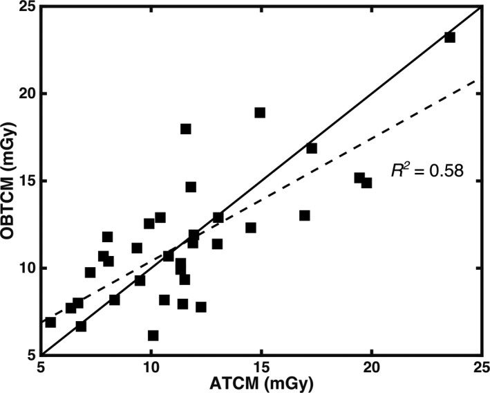
Methods: Thirty-four patients (17 females, 17 males) who underwent clinically indicated CT chest/abdomen/pelvis (CAP) examinations employing OBTCM were collected from two multi-detector row CT scanners. Patient size metric was assessed as water equivalent diameter (Dw ) taken at the center of the scan volume. Breast and lung tissues were segmented from patient image data to create voxelized models for use in a Monte Carlo transport code. The OBTCM schemes for the chest portion were extracted from the raw projection data. ATCM schemes were estimated using a recently developed method. Breast and lung doses for each TCM scenario were estimated for each patient model. CTDIvol -normalized breast (nDbreast ) and lung (nDlung ) doses were subsequently calculated. The differences between OBTCM and ATCM normalized organ dose estimates were tested using linear regression models that included CT scanner and Dw as covariates.
Results: Mean dose reduction from OBTCM in nDbreast was significant after adjusting for the scanner models and patient size (P = 0.047). When pooled with females and male patient, mean dose reduction from OBTCM in nDlung was observed to be trending after adjusting for the scanner model and patient size (P = 0.085).
Conclusions: One specific manufacturer's OBTCM was analyzed. OBTCM was observed to significantly decrease normalized breast relative to a modeled version of that same manufacturer's ATCM scheme. However, significant dose savings were not observed in lung dose over all. Results from this study support the use of OBTCM chest protocols for females only.
Keywords: breast and lung dose; organ-based modulation; tube current modulation.
© 2021 The Authors. Journal of Applied Clinical Medical Physics published by Wiley Periodicals, Inc. on behalf of American Association of Physicists in Medicine.
Conflict of interest statement
M McNitt‐Gray: Departmental master research agreement, Siemens Healthineers, Forchheim, Germany; Research grant support, Siemens Healthineers, Forchheim, Germany; Member, Scientific Advisory Board, Hura Imaging, LLC, Los Angeles, CA. R Layman: Research agreement, Siemens Healthineers, Forchheim, Germany; research support from National Aeronautics and Space Administration and United States Department of Agriculture, Houston, TX.
Figures


References
-
- Brenner DJ, Hall EJ. Computed tomography–an increasing source of radiation exposure. N Engl J Med. 2007;357:2277–2284. - PubMed
-
- Hopper KD, King SH, Lobell ME, et al. The breast: in‐plane x‐ray protection during diagnostic thoracic CT–shielding with bismuth radioprotective garments. Radiology. 1997;205:853–858. - PubMed
-
- Yilmaz MH, Albayram S, Yaşar D, et al. Female breast radiation exposure during thorax multidetector computed tomography and the effectiveness of bismuth breast shield to reduce breast radiation dose. J Comput Assist Tomogr. 2007;31:138–142. - PubMed
MeSH terms
Grants and funding
LinkOut - more resources
Full Text Sources
Other Literature Sources
Medical
Miscellaneous
