. 2021 Aug:70:102379.
doi: 10.1016/j.anaerobe.2021.102379. Epub 2021 Apr 30.

A cortex-specific penicillin-binding protein contributes to heat resistance in Clostridioides difficile spores

Affiliations

A cortex-specific penicillin-binding protein contributes to heat resistance in Clostridioides difficile spores

Yasir Adil Jabbar Alabdali et al. Anaerobe. 2021 Aug.

Abstract

Background: Sporulation is a complex cell differentiation programme shared by many members of the Firmicutes, the end result of which is a highly resistant, metabolically inert spore that can survive harsh environmental insults. Clostridioides difficile spores are essential for transmission of disease and are also required for recurrent infection. However, the molecular basis of sporulation is poorly understood, despite parallels with the well-studied Bacillus subtilis system. The spore envelope consists of multiple protective layers, one of which is a specialised layer of peptidoglycan, called the cortex, that is essential for the resistant properties of the spore. We set out to identify the enzymes required for synthesis of cortex peptidoglycan in C. difficile.

Methods: Bioinformatic analysis of the C. difficile genome to identify putative homologues of Bacillus subtilis spoVD was combined with directed mutagenesis and microscopy to identify and characterise cortex-specific PBP activity.

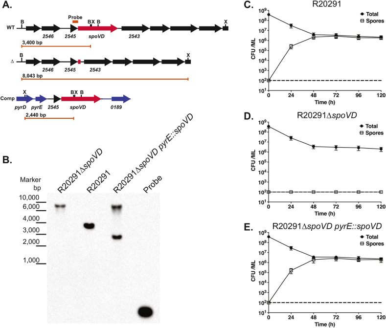
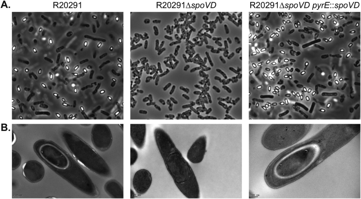
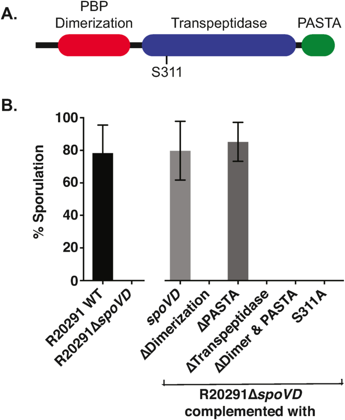
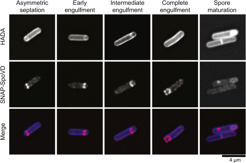
Results: Deletion of CDR20291_2544 (SpoVDCd) abrogated spore formation and this phenotype was completely restored by complementation in cis. Analysis of SpoVDCd revealed a three domain structure, consisting of dimerization, transpeptidase and PASTA domains, very similar to B. subtilis SpoVD. Complementation with SpoVDCd domain mutants demonstrated that the PASTA domain was dispensable for formation of morphologically normal spores. SpoVDCd was also seen to localise to the developing spore by super-resolution confocal microscopy.

Conclusions: We have identified and characterised a cortex specific PBP in C. difficile. This is the first characterisation of a cortex-specific PBP in C. difficile and begins the process of unravelling cortex biogenesis in this important pathogen.

Keywords: Clostridioides difficile; Penicillin-binding protein; Peptidoglycan; SpoVD; Sporulation.


Conflict of interest statement

Declaration of competing interest The authors declare that the research was conducted in the absence of any commercial or financial relationships that could be construed as a potential conflict of interest.

Figures

Fig. 1
Fig. 1
Sporulation requires SpoVDCd. A. Genomic organisation of the native spoVDCd locus (WT), following deletion of the spoVDCd gene (Δ) and following complementation by insertion of R20291_2545 and spoVDCd between the pyrE and R20291_0189 genes (Comp). The locations of XmnI (X) and BsrGI (B) sites are indicated, as is the annealing site of the Southern blot probe. The length of the diagnostic restriction product containing the probe sequence is also shown below each locus diagram. B. Southern blot analysis of a spoVDCd mutant (R20291ΔspoVD), the wild type parental strain (R20291) and complemented strain (R20291ΔspoVD pyrE:spoVD). A DNA ladder is shown on the left hand side. The predicted fragment sizes and annealing site of the probe are shown in panel A. C.-E. Sporulation efficiencies of the wild type (C.), spoVDCd mutant (D.) and complemented strains (E.). Stationary phase cultures were incubated anaerobically for 5 days with samples taken daily to enumerate total colony forming units (CFUs) and spores, following heat treatment to kill vegetative cells. Experiments were performed in duplicate on biological triplicates with mean and standard deviation shown. The dotted horizontal line indicates the limit of detection of the experiment.
Fig. 2
Fig. 2
Microscopic analysis of sporulation. Phase-contrast light microscopy (A.) and negative stained TEM (B.) of the wild type parental strain (R20291), spoVDCd mutant (R20291ΔspoVD) and complemented strain (R20291ΔspoVD pyrE:spoVD). A. Cultures were imaged at day 5 of the sporulation assays shown in Fig. 1. Spores are visible as ovoid phase bright objects i, while vegetative cells are phase dark bacilli. B. TEM imaging of developing spores clearly shows normal spore development in R20291 and R20291ΔspoVD pyrE:spoVD; the densely stained core surrounded by a thick, largely unstained cortex layer. Cultures of R20291ΔspoVD contained no morphologically normal developing spores, although fully engulfed prespores without a cortex (example shown) were common.
Fig. 3
Fig. 3
The contribution of SpoVDCd domains to sporulation. A. The domain organisation of SpoVDCd showing Pfam predictions [33]. B. Sporulation efficiency of R20291, R20291ΔspoVD and R20291ΔspoVD complemented in trans using plasmids expressing a series of mutant CLIP-SpoVDs under the control of a constitutive promoter: full-length CLIP-SpoVDCd (spoVD); CLIP-SpoVDCd lacking the PBP dimerization domain (ΔDimerization), PASTA domain (ΔPASTA), transpeptidase domain (ΔTranspeptidase) or both PBP dimerization and PASTA domains (ΔDimer & PASTA); CLIP-SpoVDCd lacking the active site nucleophile serine (S311A). Shown is the sporulation efficiency after 5 days in broth culture, expressed as number of spores as a percentage of total viable CFUs. Experiments were conducted in duplicate on biological triplicates and mean and standard deviations are shown.
Fig. 4
Fig. 4
Subcellular localisation of SpoVDCd. R20291 SNAP-spoVDCd was grown for 24 h in TY broth containing the fluorescent d-amino acid HADA (500 nM) to label de novo synthesised peptidoglycan. The bacteria were then further stained with SNAP-Cell TMR-Star (250 nM) to label SNAP-SpoVDCd, fixed, mounted in SlowFade Diamond mountant and imaged using a Zeiss AiryScan confocal microscope. Shown are representative cells demonstrating the sequential stages of sporulation: asymmetric septum placement, early, intermediate and complete prespore engulfment respectively, and spore maturation.

References

    1. Lessa F.C., Mu Y., Bamberg W.M., Beldavs Z.G., Dumyati G.K., Dunn J.R. Burden of Clostridium difficile infection in the United States. N. Engl. J. Med. 2015;372:825–834. - PMC - PubMed
    1. Smits W.K., Lyras D., Lacy D.B., Wilcox M.H., Kuijper E.J. Clostridium difficile infection. Nat Rev Dis Primers. 2016;2:16020. - PMC - PubMed
    1. Rupnik M., Wilcox M.H., Gerding D.N. Clostridium difficile infection: new developments in epidemiology and pathogenesis. Nat. Rev. Microbiol. 2009;7:526–536. - PubMed
    1. He M., Miyajima F., Roberts P., Ellison L., Pickard D.J., Martin M.J. Emergence and global spread of epidemic healthcare-associated Clostridium difficile. Nat. Genet. 2013;45:109–113. - PMC - PubMed
    1. Deakin L.J., Clare S., Fagan R.P., Dawson L.F., Pickard D.J., West M.R. The Clostridium difficile spo0A gene is a persistence and transmission factor. Infect. Immun. 2012;80:2704–2711. - PMC - PubMed

MeSH terms

Substances