Review
. 2021 May 3;20(1):44.
doi: 10.1186/s12944-021-01466-0.

Sphingolipid lysosomal storage diseases: from bench to bedside

Affiliations
Review

Sphingolipid lysosomal storage diseases: from bench to bedside

Muna Abed Rabbo et al. Lipids Health Dis. .

Abstract

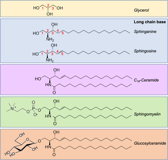
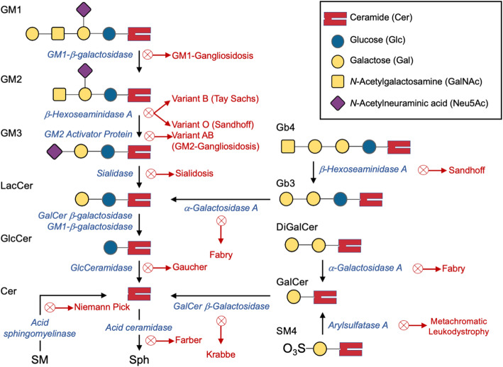
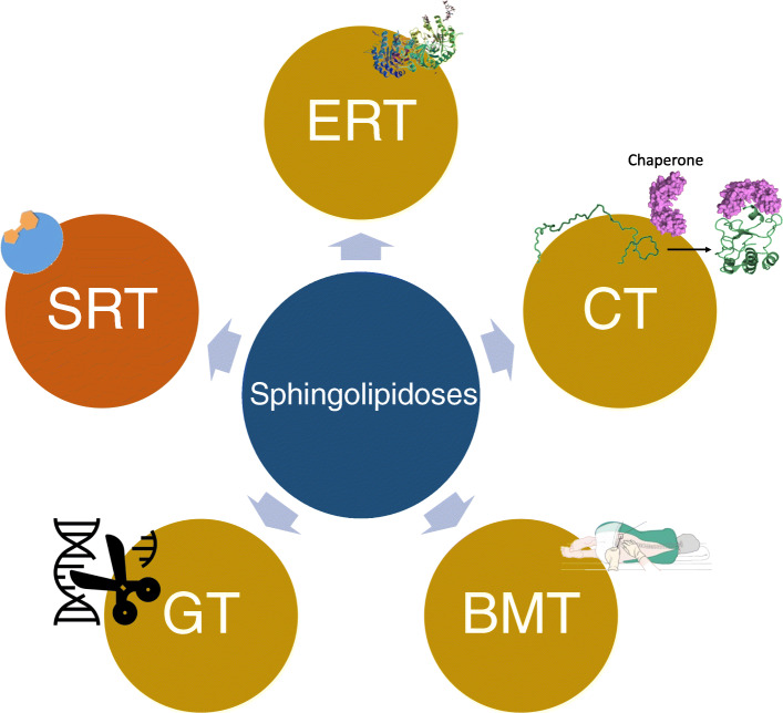
Johann Ludwig Wilhelm Thudicum described sphingolipids (SLs) in the late nineteenth century, but it was only in the past fifty years that SL research surged in importance and applicability. Currently, sphingolipids and their metabolism are hotly debated topics in various biochemical fields. Similar to other macromolecular reactions, SL metabolism has important implications in health and disease in most cells. A plethora of SL-related genetic ailments has been described. Defects in SL catabolism can cause the accumulation of SLs, leading to many types of lysosomal storage diseases (LSDs) collectively called sphingolipidoses. These diseases mainly impact the neuronal and immune systems, but other systems can be affected as well. This review aims to present a comprehensive, up-to-date picture of the rapidly growing field of sphingolipid LSDs, their etiology, pathology, and potential therapeutic strategies. We first describe LSDs biochemically and briefly discuss their catabolism, followed by general aspects of the major diseases such as Gaucher, Krabbe, Fabry, and Farber among others. We conclude with an overview of the available and potential future therapies for many of the diseases. We strive to present the most important and recent findings from basic research and clinical applications, and to provide a valuable source for understanding these disorders.

Keywords: Fabry; Gaucher; Krabbe; gangliosidosis; inborn errors of metabolism; lysosomal storage diseases; neurological diseases; sphingolipidoses; sphingolipids.


Conflict of interest statement

The authors declare that they have no competing interests.

Figures

Fig. 1
Fig. 1
Common structures of representative SLs. SLs contain a long-chain base whether sphinganine (saturated) or sphingosine (monounsaturated at C4). N-acylation at the amino group on C2 creates ceramide, which may consist of varying number of carbon atoms. The representative ceramide shown here is palmitoyl-ceramide that contains 16 carbons. The addition of a phosphocholine at C1 creates the parent phosphosphingolipid sphingomyelin, and glycosylation at the same carbon generates glucosylceramide
Fig. 2
Fig. 2
Three general pathways for the generation of Cer. In mammalian cells, Cer is biosynthesized de novo or generated by catabolism of complex SLs. In the de novo synthesis pathway (purple block arrow), a four-enzyme sequence culminates in the formation of Cer from the amino acid serine and palmitoyl-CoA. This pathway is located in the ER. Sphingomyelin can be hydrolyzed to Cer in the SM hydrolysis pathway (orange block arrow), which is a one-enzyme step. (Degradation of other complex SLs is not shown.) Alternatively, Cer can be produced in the salvage pathway (green block arrows), through the acylation of Sph by the ceramide synthase family of enzymes. The red blocks represent cartoons of the possible structures of the molecules. It should be noted that the acyl-chain length of Cer can vary greatly
Fig. 3
Fig. 3
Lysosomal SL catabolism and enzyme deficiencies causing storage diseases. A schematic of the various SL metabolic pathways is presented, indicating the enzymes whose deficiency leads to several diseases. Note that each enzyme is assisted by one or more Saps: GM2A assists both β-gal and β-hexosaminidase. SapB assists sialidase, α-GAL, GALC, and β-gal. SapC assists GALC, β-gal, GCase, acid ceramidase, and GALC. Acid ceramidase is also assisted by SapD
Fig. 4
Fig. 4
Summary of the major cellular interactions leading to the neurological features of sphingolipidoses. A schematic of the main events that lead to caspase-dependent and independent activation of neuronal apoptosis through myriad intercalated pathways, such as the disruption of TFEEB fine-tuning, impaired Ca2+ homeostasis in smooth ER, mitochondria and lysosomes, lysosomal membrane permeabilization, and impaired autophagy, along with others (not depicted here). Proteins are depicted in oblong shapes, while lipids are in circles and cellular events are in rectangles. Upward green arrows represent increase in cellular concentration while downward red arrows illustrate decreased concentration. Note that organellar sizes are not to scale. not to scale. This figure was created in Biorender.com
Fig. 5
Fig. 5
General therapeutic strategies for the treatment of sphingolipidoses. There are five main therapeutic approaches to treat sphingolipidoses. Substrate reduction therapy (SRT, orange) involves the prevention of influx of substrates into the lysosome, to lower synthesis of the accumulating substance. The other treatment strategies (gold) involve enhancing the activity of the missing or malfunctioning enzyme. Enzyme replacement therapy (ERT) uses purified enzyme to reverse the pathology (the crystal structure of α-GAL is shown). Chaperone therapy (CT) is used to assist the folding of misfolded enzymes to be targeted to the lysosome. Bone marrow transplantation and stem cell transplantation (BMT) are used to supply the body with the correct form of the missing enzyme, and gene therapy (GT) is used to modify the genes responsible for the aberrant phenotype

References

    1. Kolter T, Sandhoff K. Lysosomal degradation of membrane lipids. FEBS Lett. 2010;584:1700–1712. - PubMed
    1. Lahiri S, Futerman AH. The metabolism and function of sphingolipids and glycosphingolipids. Cell Mol Life Sci. 2007;64:2270–2284. - PMC - PubMed
    1. Abou-Ghali M, Stiban J. Regulation of ceramide channel formation and disassembly: Insights on the initiation of apoptosis. Saudi J Biol Sci. 2015;22:760–772. - PMC - PubMed
    1. Albeituni S, Stiban J. Roles of Ceramides and Other Sphingolipids in Immune Cell Function and Inflammation. Adv Exp Med Biol. 2019;1161:169–191. - PubMed
    1. Futerman A. Biochemistry of Lipids, Lipoproteins and Membranes 6th Edition. 2016. Sphingolipids; pp. 297–326.