. 2021 Oct;11(10):2564-2581.
doi: 10.1158/2159-8290.CD-20-1540. Epub 2021 May 3.

Inhibition of CDK4/6 Promotes CD8 T-cell Memory Formation

Affiliations

Inhibition of CDK4/6 Promotes CD8 T-cell Memory Formation

Max Heckler et al. Cancer Discov. 2021 Oct.

Abstract

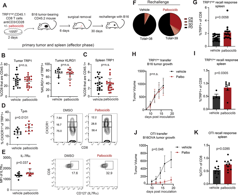
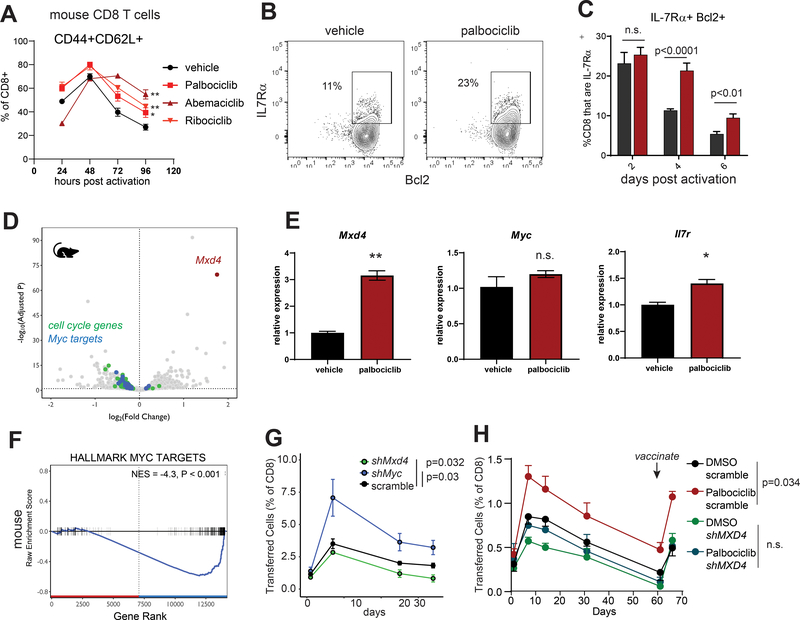
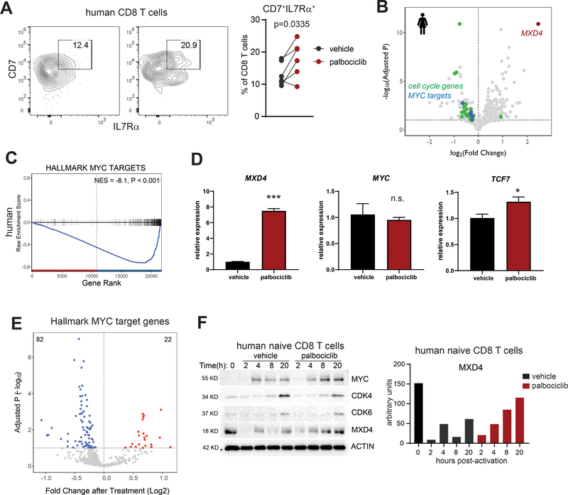
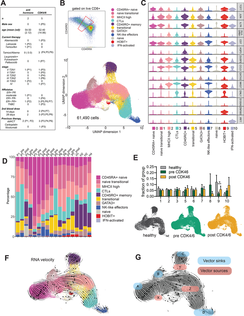
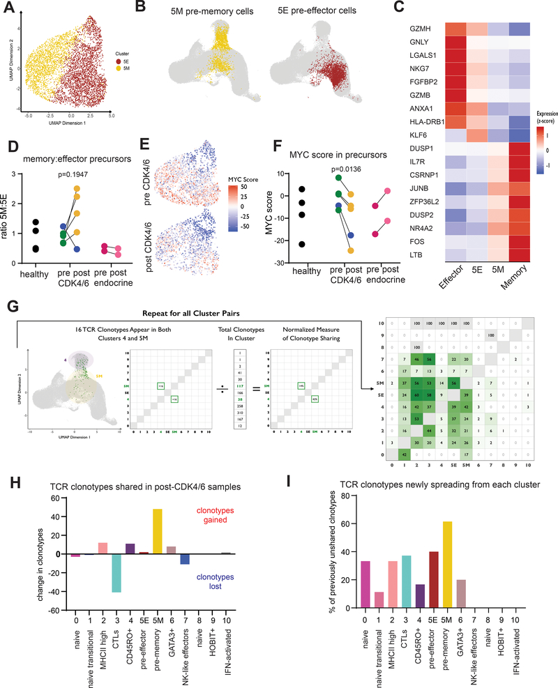
CDK4/6 inhibitors are approved to treat breast cancer and are in trials for other malignancies. We examined CDK4/6 inhibition in mouse and human CD8+ T cells during early stages of activation. Mice receiving tumor-specific CD8+ T cells treated with CDK4/6 inhibitors displayed increased T-cell persistence and immunologic memory. CDK4/6 inhibition upregulated MXD4, a negative regulator of MYC, in both mouse and human CD8+ T cells. Silencing of Mxd4 or Myc in mouse CD8+ T cells demonstrated the importance of this axis for memory formation. We used single-cell transcriptional profiling and T-cell receptor clonotype tracking to evaluate recently activated human CD8+ T cells in patients with breast cancer before and during treatment with either palbociclib or abemaciclib. CDK4/6 inhibitor therapy in humans increases the frequency of CD8+ memory precursors and downregulates their expression of MYC target genes, suggesting that CDK4/6 inhibitors in patients with cancer may augment long-term protective immunity. SIGNIFICANCE: CDK4/6 inhibition skews newly activated CD8+ T cells toward a memory phenotype in mice and humans with breast cancer. CDK4/6 inhibitors may have broad utility outside breast cancer, particularly in the neoadjuvant setting to augment CD8+ T-cell priming to tumor antigens prior to dosing with checkpoint blockade.This article is highlighted in the In This Issue feature, p. 2355.


Figures

Figure 1:
Figure 1:. Treatment of tumor-specific CD8 T cells ex vivo with CDK4/6i induces long-term protective memory in mice.
A) Diagram of experimental protocol. CD8 T cells were isolated by magnetic beads from pooled spleen and LNs of TRP1high CD45.1+ mice and activated with anti-CD3/28 beads in the presence of vehicle (DMSO) or 500nM palbociclib for 48 hours. Cells were washed and transferred into CD45.2+ mice bearing 100mm3 B16 tumors. Tumors were surgically removed after 6 days, and the mice recovered for 30 days prior to inoculation with 500,000 B16 cells subcutaneously on the opposite flank. B) Surgically excised tumors were digested and analyzed by flow cytometry for the frequency of CD45.1+ TRP1 cells and their expression of KLRG1. C) Spleens were analyzed by flow cytometry from mice 6 days after transfer of activated TRP1high cells. D) Surgically excised tumors from A were digested and analyzed by flow cytometry. Peripheral memory (Tpm) cells were defined as CD45.1+ CX3CR1intermediate as shown in the representative flow plots. Data pooled from 2 independent experiments. E) IL-7Rα expression on tumor-infiltrating TRP1high cells at the time of surgery. Representative flow plots are shown. Data pooled from 2 independent experiments. F) Pie charts show the response to B16 rechallenge pooled from 5 independent experiments by defining tumor response on the day that the first tumor reached 1000mm3 (day 12–16 post inoculation). complete response (CR, <10 mm3); partial response (PR, <200 mm3); stable disease (SD, <800 mm3); and progressive disease (<800 mm3). G) Frequency of TRP1high CD45.1+ cells as a fraction of total CD8 T cells in spleens of mice 10 days after tumor rechallenge. Data pooled from 3 independent experiments. H) TRP1low CD45.2 CD8 T cells were activated in vitro for 48 hours with peptide-pulsed antigen presenting cells in the presence of palbociclib or vehicle and transferred into CD45.1+ mice bearing B16 tumors according to the scheme in panel A. Upon tumor rechallenge, tumor growth was similar between vehicle and palbociclib groups. N=10 per group. I) Frequency of TRP1low CD45.2+ cells as a fraction of total CD8 T cells in spleens of mice 17 days after tumor rechallenge. J) OT-I CD8 T cells were activated in vitro for 48 hours with anti-CD3/CD28 beads in the presence of palbociclib or vehicle and transferred into C57BL/6 mice bearing B16OVA tumors according to the scheme in panel A. Tumor growth during the rechallenge phase is shown. N=10 per group. K) Frequency of SIINFEKL tetramer+ CD8 T cells as a fraction of total CD8 T cells in spleens of mice 19 days after tumor rechallenge. Error bars are SEM throughout.
Figure 2:
Figure 2:. CDK4/6 inhibitors induce long term persistence of CD8 T cells independent of cell cycle.
A) Naïve CD8 T cells were labeled with CFSE and activated with anti-CD3/CD28 beads in the presence of 500nM palbociclib, 1μM AuroraKi, 100nM CDK7i, 150nM PLKi, 4.5 μM CDK1/2i, or vehicle. Proliferation indexes were calculated based on number of cell divisions determined by flow cytometry after 4 days. B) Diagram of experimental protocol. TRP1low CD45.2+ CD8 T cells were activated in the presence of the indicated cell cycle inhibitors at the concentrations used in A and transferred into CD45.1 recipients. Frequency of transferred cells in peripheral blood was measured over time. Representative of 3 independent experiments. C) At 85 days post transfer, the mice in B were immunized with irradiated B16-GVAX admixed with TRP1 peptide. Frequency of TRP1low cells was analyzed in the spleen 6 days later. Representative of 3 independent experiments. D) Polyclonal CD45.2+ CD8 T cells were activated in the presence of the indicated CDK4/6 inhibitors and transferred into CD45.1 recipients. Frequency of transferred cells in peripheral blood was measured over time. Representative of 3 independent experiments. E) After 56 days, mice from D were analyzed for frequencies of transferred cells in spleen and in blood. Plot shows correlation between the two compartments. Individual mice are represented by colored dots representing the treatment groups. F) Polyclonal CD45.2+ CD8 T cells were stained with CFSE and activated in vitro with anti-CD3/28 beads. Fast or slow cycling cells were sorted by FACS using the indicated gates, transferred into CD45.1+ recipients and monitored over time in peripheral blood. Error bars are SEM throughout.
Figure 3:
Figure 3:. CDK4/6 inhibition acts through the Mxd4/Myc transcription factor axis to inducea memory precursor phenotype mouse CD8 T cells.
A) CD8 T cells were isolated by magnetic beads from pooled spleen and LNs of a C57BL/6 mouse and activated in vitro with anti-CD3/28 beads in the presence of the indicated compounds. Cells were analyzed by flow cytometry for expression of CD44 and CD62L at the indicated times. Representative of 3 independent experiments. B) CD8 T cells were isolated by negative selection on magnetic beads from pooled spleen and lymph nodes of a C57BL/6 mouse. Cells were labeled with CFSE and activated with anti-CD3/CD28 beads in the presence of the indicated compounds. 72 hours later cells were analyzed by flow cytometry. C) Quantification of Bcl-2 and IL7Rα by flow cytometry after gating on cells experiencing at least one cell division as shown in B. D) Murine TRP1high CD8 T cells were activated for 48 hours with anti-CD3/28 beads in the presence of vehicle or 500nM palbociclib. Differentially expressed transcripts were identified by bulk RNAseq. Hallmark cell cycle genes are shown in green; hallmark Myc targets are shown in blue. E) Quantitative PCR validation of the indicated transcript levels in mouse CD8 T cells at 48 hours post activation. Representative of 2 independent experiments. *p<0.05, **p<0.01. Error bars are SEM. F) Gene-set enrichment analysis of MYC target genes in mouse palbociclib treated CD8 T cells. G) TRP1high CD45.1+ CD8 T cells were transduced with retroviruses encoding RFP and scrambled, Mxd4 or Myc shRNAs. Cells were transferred into CD45.2+ recipient mice, and their frequency in peripheral blood was monitored over time. Representative of 2 independent experiments. Area under the curve values were calculated for individual mice and p values determined versus the scrambled control group using a Mann-Whitney test. H) Similar to G, TRP1 CD8 T cells were silenced and activated in the presence of vehicle or palbociclib. After 60 days, mice were vaccinated with TRP1 peptide mixed with irradiated B16-GVAX.
Figure 4:
Figure 4:. CDK4/6 inhibition and MXD4-Myc transcription factors regulate memory cell fate decisions in human CD8 T cells.
A) CD8 T cells were isolated by magnetic beads from peripheral blood of healthy donors and activated in vitro with anti-CD3/28 beads in the presence of vehicle or palbociclib. Cells were analyzed by flow cytometry for expression of IL7Rα and CD7 at 48 hours. Matched pairs are from six individual donors. Data pooled from 2 independent experiments. B) Human CD8 T cells were isolated by magnetic beads from healthy blood donors and activated in vitro with anti-CD3/28 beads in the presence of vehicle, abemaciclib or palbociclib. RNAseq was performed on 48-hour samples. Differential expression analysis was performed between vehicle treated and combined CDK4/6i treated cells. C) Gene-set enrichment analysis of MYC target genes in human CDK4/6i treated CD8 T cells. D) Quantitative PCR validation of the indicated transcript levels in human CD8 T cells at 48 hours post activation. Representative of 2 independent experiments. *p<0.05, ***p<0.001. Error bars are SEM. E) CD8 T cells were isolated by negative selection from the blood of healthy donors and treated for 48 hours with palbociclib, abemaciclib, or vehicle. Areas of open, transcriptionally active chromatin were identified and grouped into consensus peaks. Peaks corresponding to open chromatin were observed in 184 genes (92%) from the hallmark MYC target gene set. The change in accessibility at each peak after treatment with palbociclib is shown as a volcano plot. P values were FDR-controlled. F) Human naïve CD8 T cells were isolated by magnetic beads and activated with anti-CD3/CD28 beads for the indicated times. Protein lysates were prepared from 3 healthy donors, pooled, and analyzed by immunoblot for the indicated proteins. MXD4 protein levels were quantified. Representative of 2 independent experiments.
Figure 5:
Figure 5:. Memory cell precursors can be found by single cell transcriptional analysis of recently activated CD8 T cells from human peripheral blood.
A) Clinical characteristics of the patient cohort. B) Peripheral blood mononuclear cells were enriched for CD8 T cells using magnetic beads and stained with antibodies to CD8, CD45RO, and CD45RA. Cells were gated on live CD8+ prior to sorting the population gated in red. Single cell transcriptional profiling was performed using the 10X Genomics platform. 61,889 cells were analyzed from a combined 4 healthy donors and paired pre- and on-treatment samples from the 7 breast cancer patients shown in A. Cells were projected onto 2 dimensions by UMAP and grouped into 11 clusters. C) Violin plots of select cluster defining genes. D) Cluster representation per sample. E) Fractional representation of healthy, pre CDK4/6 and post CDK4/6 cells represented in each cluster. CD45RA+ naïve cells (cluster 0) and CD45RO+ memory cells (cluster 4) were excluded from the analysis. Error bars are SEM. Significance was evaluated using paired analysis of pre-CDK4/6 and post-CDK4/6 values for each patient; only cluster 9 was significant, *p=0.0403. UMAP projections of cells from healthy, pre and post CDK4/6 samples are shown. P1 and P2 post samples are included in the pre-CDK4/6 plot. F) RNA velocity analysis. G) Diagram of sinks and sources based on RNA velocity.
Figure 6:
Figure 6:. TCR clonotype tracing reveals a transitional population of pre-effector and pre-memory cells in peripheral blood whose fractional composition and MYC signature are affected by CDK4/6i treatment.
A) Cluster 5 (transitional cells) was further divided into 2 sub-clusters. B) UMAP projections of sub-clusters 5M and 5E. C) Top differentially expressed genes among cluster 5 sub-clusters are displayed as a heatmap. Expression scores for those same genes are shown for memory cells (Cluster 4) and cytolytic effector cells (Cluster 3). D) Ratio of cells in sub-clusters 5M:5E analyzed in pairwise fashion. Blue dots indicate P5 (dominant clonotype has influenza-specific TCR). E) MYC scores for individual cells in Cluster 5. F) MYC hallmark gene signature was defined across the entire dataset and analyzed in pairwise comparison between pre and on CDK4/6 inhibitor treatment and between pre- and on-treatment samples from control patients receiving endocrine therapy. G) TCR clonotypes that were shared across clusters in pre-CDK4/6 samples are plotted in the matrix shown in green as percent of total clonotypes in that cluster. H) For each cluster, the total number of shared clonotypes gained was subtracted from the total number of shared clonotypes lost to quantify the total flux of TCR clonotypes occurring for each cluster. I) For each cluster, the number of unshared clonotypes in the pre-treatment samples was identified. Of these, clonotypes that became shared in the on-treatment samples are plotted as a percentage of total previously unshared for each cluster.

References

    1. Dougan MD,G; Dougan SK Cancer Immunotherapy: Beyond Checkpoint Blockade. Annual Review of Cancer Biology. 2019;3(1):55–75. - PMC - PubMed
    1. Huang AC, Postow MA, Orlowski RJ, Mick R, Bengsch B, Manne S, et al.T-cell invigoration to tumour burden ratio associated with anti-PD-1 response. Nature. 2017;545(7652):60–5. - PMC - PubMed
    1. Pedicord VA, Montalvo W, Leiner IM, and Allison JP. Single dose of anti-CTLA-4 enhances CD8+ T-cell memory formation, function, and maintenance. Proc Natl Acad Sci U S A. 2011;108(1):266–71. - PMC - PubMed
    1. Hodi FS, O’Day SJ, McDermott DF, Weber RW, Sosman JA, Haanen JB, et al.Improved survival with ipilimumab in patients with metastatic melanoma. N Engl J Med. 2010;363(8):711–23. - PMC - PubMed
    1. Kaech SM, and Ahmed R. Memory CD8+ T cell differentiation: initial antigen encounter triggers a developmental program in naive cells. Nat Immunol. 2001;2(5):415–22. - PMC - PubMed

Publication types

MeSH terms