. 2021 May 11;118(19):e2016146118.
doi: 10.1073/pnas.2016146118.

Reversed-engineered human alveolar lung-on-a-chip model

Affiliations

Reversed-engineered human alveolar lung-on-a-chip model

Di Huang et al. Proc Natl Acad Sci U S A. .

Abstract

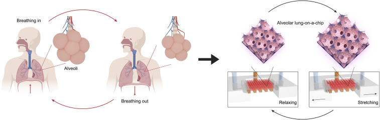
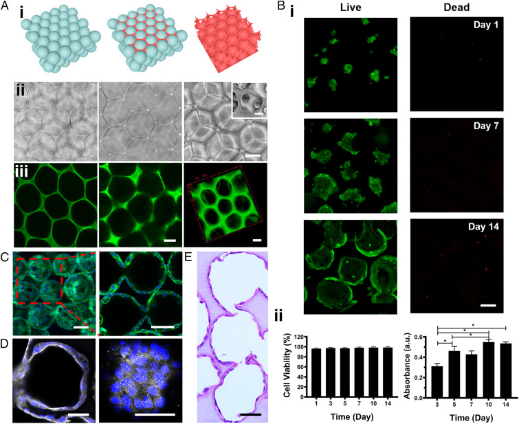
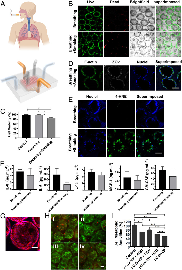
Here, we present a physiologically relevant model of the human pulmonary alveoli. This alveolar lung-on-a-chip platform is composed of a three-dimensional porous hydrogel made of gelatin methacryloyl with an inverse opal structure, bonded to a compartmentalized polydimethylsiloxane chip. The inverse opal hydrogel structure features well-defined, interconnected pores with high similarity to human alveolar sacs. By populating the sacs with primary human alveolar epithelial cells, functional epithelial monolayers are readily formed. Cyclic strain is integrated into the device to allow biomimetic breathing events of the alveolar lung, which, in addition, makes it possible to investigate pathological effects such as those incurred by cigarette smoking and severe acute respiratory syndrome coronavirus 2 pseudoviral infection. Our study demonstrates a unique method for reconstitution of the functional human pulmonary alveoli in vitro, which is anticipated to pave the way for investigating relevant physiological and pathological events in the human distal lung.

Keywords: alveoli; distal lung; inverse opal; lung-on-a-chip; three-dimensional.


Conflict of interest statement

The authors declare no competing interest.

Figures

Fig. 1.
Fig. 1.
The breathing human alveolar lung-on-a-chip. Schematics showing the distal lung, the breathing cycles, and the in vitro on-chip model of the breathing alveolar lung.
Fig. 2.
Fig. 2.
Fabrication of the alveoli-like 3D GelMA inverse opal structure and formation of the alveolar lung model. (A, i and ii) Schematics and bright-field optical images showing the fabrication process of the alveoli-like 3D GelMA inverse opal structure. Alginate microbeads with uniform sizes (201 ± 12 µm) were first assembled into a cubic close-packed lattice (Left); a 7% (wt/wt) GelMA solution was then infiltrated into the void spaces of the lattice and cross-linked (Center). Finally, the alginate microbeads were selectively removed using EDTA, leaving behind an alveoli-like hydrogel structure (Right). (Scale bar: 100 µm.) A, ii, Right, Inset shows an scanning electron microscopy image of the interconnecting window between two adjacent sacs. (Scale bar: 20 µm; note the shrinkage of the sac size comparing to optical images was caused by the sample-drying process for scanning electron microscopy imaging.) (A, iii) Fluorescence confocal images illustrating the GelMA inverse opal hydrogel structure, where GelMA was chemically labeled with FITC. (Left) After infiltration of the GelMA solution into the void spaces of the alginate microbead lattice and cross-linking, (Center) after removal of the alginate microbeads, and (Right) 3D reconstruction of the GelMA hydrogel structure. (Scale bars: 100 µm.) (B, i) Fluorescence confocal images showing the hAECs cultured in the GelMA inverse opal hydrogel structures at 1, 7, and 14 d. Green, live cells; red, dead cells. (Scale bar: 100 µm.) (B, ii) Quantification of viability (Left) and proliferation (Right) of the hAECs in the GelMA inverse opal hydrogel structures. *P < 0.05. (C) Confocal reconstruction view (Left) and sectional view (Right) of the hAECs after culturing for 14 d in the GelMA inverse opal hydrogel structure, in which the fully confluent alveolar epithelium was formed. Green, F-actin; blue, nuclei. (Scale bars: 100 µm.) (D) Confocal micrographs showing the presence of continuous tight junctions of the rim (Left) and bottom (Right) of a sac after culturing for 14 d in the GelMA inverse opal hydrogel structure. White, ZO-1; blue, nuclei. (Scale bars: 50 µm.) (E) Representative image of an H&E-stained section of the alveolar lung model after 14 d of culture. (Scale bar: 50 µm.)
Fig. 3.
Fig. 3.
Comparisons of the alveolar epithelium formation for hAECs cultured on 3D and 2D substrates of GelMA and PDMS. (A) Scanning electron microscopy images of the hAECs cultured on/in the 3D GelMA inverse opal structure (Upper) and the 2D GelMA hydrogel (Lower) at days 3, 7, and 14. (Scale bars: 25 μm.) (B) Normalized proliferation of hAECs on the two GelMA surfaces. ***P < 0.001. (C) Caspase-3/7 fluorescence signal analysis and annexin V-FITC/PI flow cytometry plots of hAECs on the two GelMA surfaces. **P < 0.01. (D) Flow cytometry analyses of annexin V-FITC– and PI-stained hAECs cells at day 14. (E) K-means clusters of the genes for (i) GO enrichment and (ii) KEGG enrichment analyses of hAECs cultured on 2D PDMS, on 2D GelMA, and in 3D GelMA for 14 d. (F) Heat maps showing differential genes expressions relating to (i) ECM–receptor interactions and (ii) TNF signaling pathway of hAECs cultured on 2D PDMS, on 2D GelMA, and in 3D GelMA for 14 d. a.u.: any unit; PI3K-Akt: phosphatidylinositol 3-kinase-protein kinase B; HIF-1α: hypoxia-inducible factor-α.
Fig. 4.
Fig. 4.
Construction of the alveolar lung-on-a-chip model. (A) Schematic representing the alveolar lung-on-a-chip (Upper) and photograph of a device without the GelMA inverse opal structure to show the underlying fluidic channels (Lower). (Scale bar: 5 mm.) (B) Fluorescence images showing the viability of the hAECs cultured in the chips without and with the breathing events. Green, live cells; red, dead cells. (Scale bar: 100 µm.) (C) Micrographs showing the expansion of the sacs under a strain of 8%. (Scale bar: 100 µm.) (D) Images of H&E-stained sections showing the epithelium formation in the chips without and with the breathing events. (Scale bar: 50 µm.) (E) ZO-1 staining showing the tight junction formation of the epithelium in the chips without and with the breathing events. White, ZO-1; blue, nuclei. (Scale bar: 50 µm.) (F) The quantified levels of secreted cytokines IL-8, IL-6, IL-1β, MCP-1, and GM-CSF by hAECs cultured in the chips without and with the breathing events. All analyses were performed at 14 d of culture.
Fig. 5.
Fig. 5.
The effects of smoking and SARS-CoV-2 pseudoviral infection on the alveolar lung-on-chip model. (A) Schematic diagrams showing the smoking alveolar lung-on-a-chip. (B) Live/dead staining of the hAECs in the alveolar lung-on-a-chip devices before and after smoking. Green, live cells; red, dead cells. (Scale bar: 100 µm.) (C) Quantification of cell viability for hAECs. Control: without breathing and smoking; breathing: with breathing but without smoking; and breathing + smoking: with breathing and smoking. (D) Confocal fluorescence images showing the effects of smoking on tight junctions of the alveolar epithelium, revealed by ZO-1 staining. White, ZO-1; blue, nuclei. (Scale bar: 100 µm.) (E) Confocal fluorescence images showing induction of oxidative stress with smoking. Green: 4-HNE; blue, nuclei. (Scale bar: 100 µm.) (F) The quantified levels of secreted cytokines IL-8, IL-6, IL-1β, MCP-1, and GM-CSF by hAECs cultured in the chips. All analyses were performed at 14 d of culture, and smoking was conducted for 75 min. (G) Fluorescence confocal images showing the expression of ACE2 receptors by hAECs in the GelMA inverse opal structures at day 14. Red, ACE2 receptors; blue, nuclei. (Scale bar: 50 µm.) (H) Fluorescence microscopic images showing the live/dead staining for hAECs in the GelMA inverse opal structures after (i) pCoV-VP infection without antiviral drugs, (ii) pCoV-VP infection in the presence of amodiaquine (5 µM), (iii) pCoV-VP infection in the presence of remdesivir (10 µM), and (iv) pCoV-VP infection in the presence of hydroxychloroquine (40 µM). Green, live cells; red, dead cells. (Scale bar: 100 µm.) (I) MTS assay showing metabolic activities of hAECs in the GelMA inverse opal structures after pCoV-VP infection in the absence and presence of antiviral drugs. *P < 0.05; **P < 0.01; ***P < 0.001.

References

    1. Papi A., Brightling C., Pedersen S. E., Reddel H. K., Asthma. Lancet 391, 783–800 (2018). - PubMed
    1. López-Campos J. L., Tan W., Soriano J. B., Global burden of COPD. Respirology 21, 14–23 (2016). - PubMed
    1. Krammer F., et al. ., Influenza. Nat. Rev. Dis. Primers 4, 3 (2018). - PMC - PubMed
    1. Mandell L. A., Niederman M. S., Aspiration pneumonia. N. Engl. J. Med. 380, 651–663 (2019). - PubMed
    1. Reid M. J. A., et al. ., Building a tuberculosis-free world: The Lancet Commission on tuberculosis. Lancet 393, 1331–1384 (2019). - PubMed

Publication types

MeSH terms

LinkOut - more resources