. 2021 May 11;118(19):e2016974118.
doi: 10.1073/pnas.2016974118.

Membrane fusion and drug delivery with carbon nanotube porins

Affiliations

Membrane fusion and drug delivery with carbon nanotube porins

Nga T Ho et al. Proc Natl Acad Sci U S A. .

Abstract

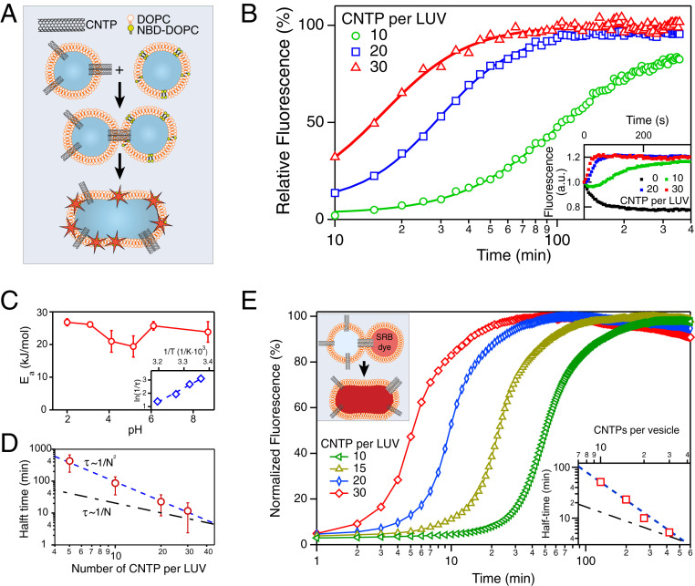
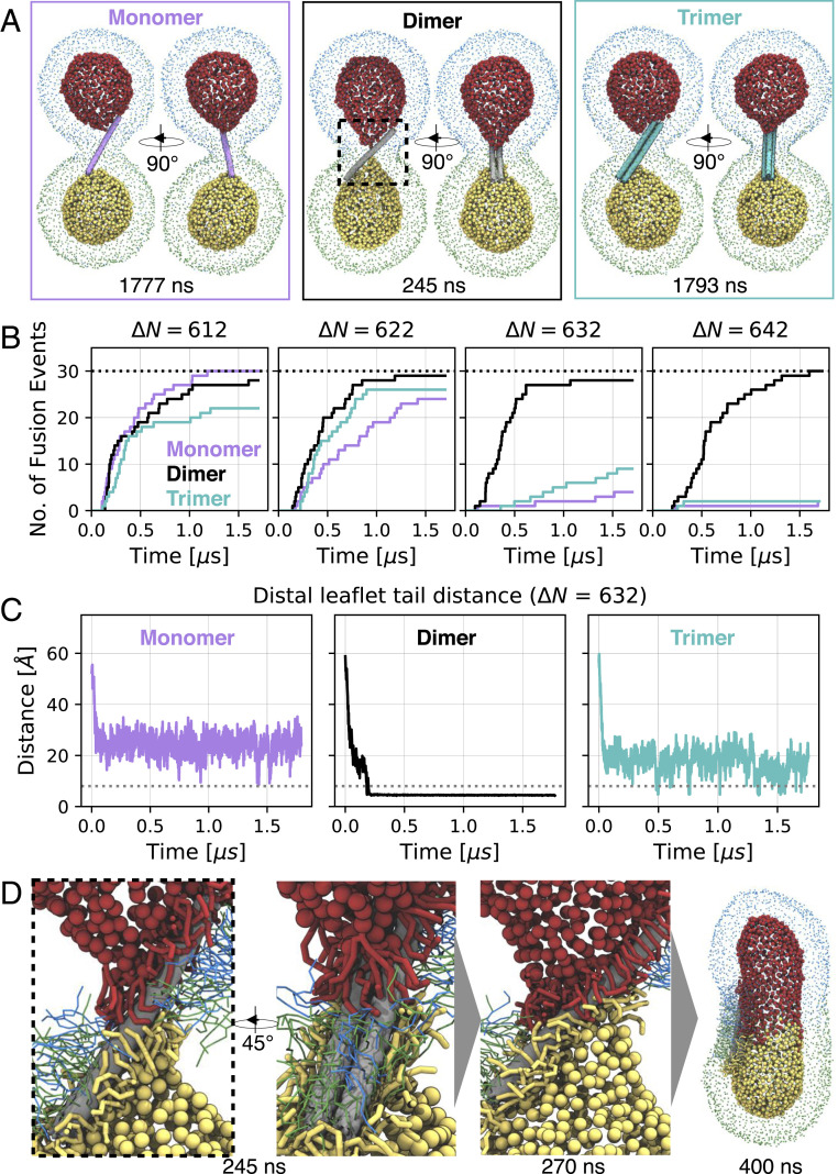
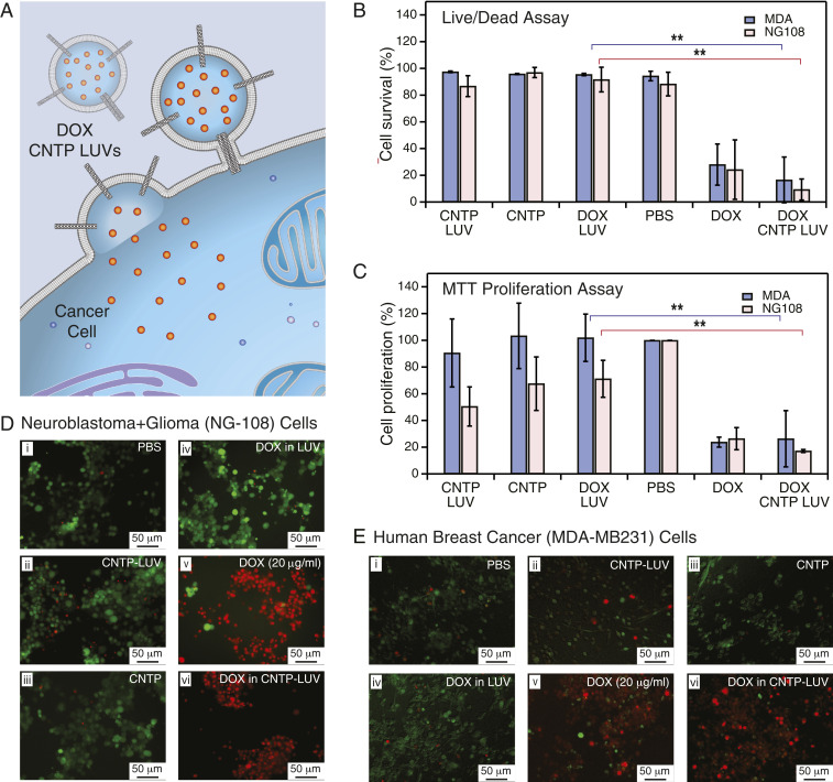
Drug delivery mitigates toxic side effects and poor pharmacokinetics of life-saving therapeutics and enhances treatment efficacy. However, direct cytoplasmic delivery of drugs and vaccines into cells has remained out of reach. We find that liposomes studded with 0.8-nm-wide carbon nanotube porins (CNTPs) function as efficient vehicles for direct cytoplasmic drug delivery by facilitating fusion of lipid membranes and complete mixing of the membrane material and vesicle interior content. Fusion kinetics data and coarse-grained molecular dynamics simulations reveal an unusual mechanism where CNTP dimers tether the vesicles, pull the membranes into proximity, and then fuse their outer and inner leaflets. Liposomes containing CNTPs in their membranes and loaded with an anticancer drug, doxorubicin, were effective in delivering the drug to cancer cells, killing up to 90% of them. Our results open an avenue for designing efficient drug delivery carriers compatible with a wide range of therapeutics.

Keywords: carbon nanotube porins; drug delivery; liposomes; membrane fusion.


Conflict of interest statement

Competing interest statement: Some of the results reported in the paper have been disclosed as part of a provisional patent filing.

Figures

Fig. 1.
Fig. 1.
CNTPs facilitate membrane fusion. (A) Schematics of the vesicle fusion assay. CNTP-LUVs fuse with the vesicles containing DOPC lipid labeled with NBD dye in self-quenching concentration, dequench the dye, and increase its fluorescence signal. (B) Kinetics of the vesicle fusion. The fluorescence intensity was recorded as NBD-LUVs were mixed with CNTP-LUVs with different average numbers of CNTPs per vesicle (as indicated on the graph). Solid lines represent best fits to the Hill equation. (C) Activation energy, Ea, for vesicle fusion plotted as a function of pH. (Inset) A representative Arrhenius plot used to obtain the Ea values (n = 3). (D) Plot of the fusion half-time as a function of the average number of CNTPs per vesicle (n = 3 for 10, 20, and 30 CNTP/LUV and n = 2 for 5 CNTP/LUV). The blue dashed line represents a fit to the second-order kinetics. The dashed-dotted black line, which corresponds to the first-order kinetics, is provided as a guide to the eye. (E) Content-mixing assay showing fluorescence signal kinetics recorded as CNTP-LUVs were exposed to LUVs encapsulating SRB dye (each curve is an average of two runs; see SI Appendix, Fig. S2 for raw traces). (Inset) The plot of the fusion half-time as a function of the average number of CNTPs per vesicle.
Fig. 2.
Fig. 2.
Coarse-grained MD simulations of CNTP-mediated vesicle fusion. (A) Snapshots of simulated systems of a CNTP monomer (magenta), dimer (gray), and trimer (cyan) (ΔN=632). Lipid phosphate groups are colored uniquely for each leaflet and vesicle (outer leaflets: blue/green for top/bottom vesicle; inner leaflets: red/yellow for top/bottom vesicle). Inner leaflet phosphate groups are drawn larger for clarity. Times of snapshots are indicated (see also Movie S1). (B) Cumulative number of CNTP-mediated vesicle fusion events as a function of time at different number asymmetries ΔN. Monomer (magenta), dimer (black), and trimer (cyan) simulations are compared. A total of 30 simulations were performed for each starting configuration (indicated as black dashed line). Simulations were 1.7 μs long. (C) Minimal distance of C5A/B tail beads of the opposing inner leaflet lipids at ΔN=632. Exemplary trace shown for monomer, dimer, and trimer, respectively. All traces for all systems are shown in SI Appendix, Fig. S6. The dashed line at 8 Å indicates contact of the opposing inner leaflets. (D) Zoom-in on CNTP dimer-mediated fusion. Time points of snapshots are indicated. Lipids within 8 Å of the CNTP are shown. Color scheme as in A. Inner leaflet lipids are drawn thicker for clarity. Outer leaflet phosphate groups are omitted for clarity (see also Movie S2).
Fig. 3.
Fig. 3.
DOX delivery with CNTPs. (A) Schematic showing CNTP-LUV loaded with the DOX payload fusing to a cancer cell and delivering DOX to the cell interior. (B) Cell survival in live/dead assay after 48-h exposure of neuroblastoma-glioma (NG-108) and human breast cancer (MDA) cell cultures to DOX-CNTP-LUVs, CNTP-LUVs, free CNTPs, DOX-LUVs, free DOX, and PBS buffer (n = 9). **P 0.01. (C) Results of MTT cell proliferation assay after 48-h exposure of NG-108 and MDA cell cultures to DOX-CNTP-LUVs, CNTP-LUVs, free CNTPs, DOX-LUVs, free DOX, and PBS buffer. (NG-108 cells: n = 9; MDA cells: n = 15). **P 0.01. (D) Fluorescence microscopy images of NG108 cell culture with live and dead cells stained with green and red dye, respectively. Prior to imaging the cells were exposed for 48 h to (i) PBS buffer, (ii) CNTP-LUVs without the drug payload, (iii) CNTP solution, (iv) LUVs encapsulating DOX, (v) 20 μg/mL of DOX, or (vi) CNTP-LUVs with encapsulated DOX. (E) Fluorescence microscopy images of MDA cell culture with live and dead cells stained with green and red dye, respectively. Prior to imaging the cells were exposed for 48 h to (i) PBS buffer, (ii) CNTP-LUVs without the drug payload, (iii) CNTP solution, (iv) LUVs encapsulating DOX, (v) 20 μg/mL of DOX, or (vi) CNTP-LUVs with encapsulated DOX.

References

    1. Katzung B. G.. Basic & Clinical Pharmacology (McGraw-Hill, New York, ed. 14, 2018).
    1. Chabner B. A., Roberts T. G., Chemotherapy and the war on cancer. Nat. Rev. Canc. 5 (1), 65–72 (2005). - PubMed
    1. Timko B. P., et al. , Advances in drug delivery. Annu. Rev. Mater. Res. 41, 1–20 (2011).
    1. Sercombe L., et al. , Advances and challenges of liposome assisted drug delivery. Front. Pharmacol. 6, 286 (2015). - PMC - PubMed
    1. Rosen Y., Gurman P., Elman N., Drug Delivery: An Integrated Clinical and Engineering Approach (CRC Press, 2017).

Publication types