. 2021 May 3;7(1):90.
doi: 10.1038/s41420-021-00465-5.

MDM2 inhibitor APG-115 exerts potent antitumor activity and synergizes with standard-of-care agents in preclinical acute myeloid leukemia models

Affiliations

MDM2 inhibitor APG-115 exerts potent antitumor activity and synergizes with standard-of-care agents in preclinical acute myeloid leukemia models

Douglas D Fang et al. Cell Death Discov. .

Abstract

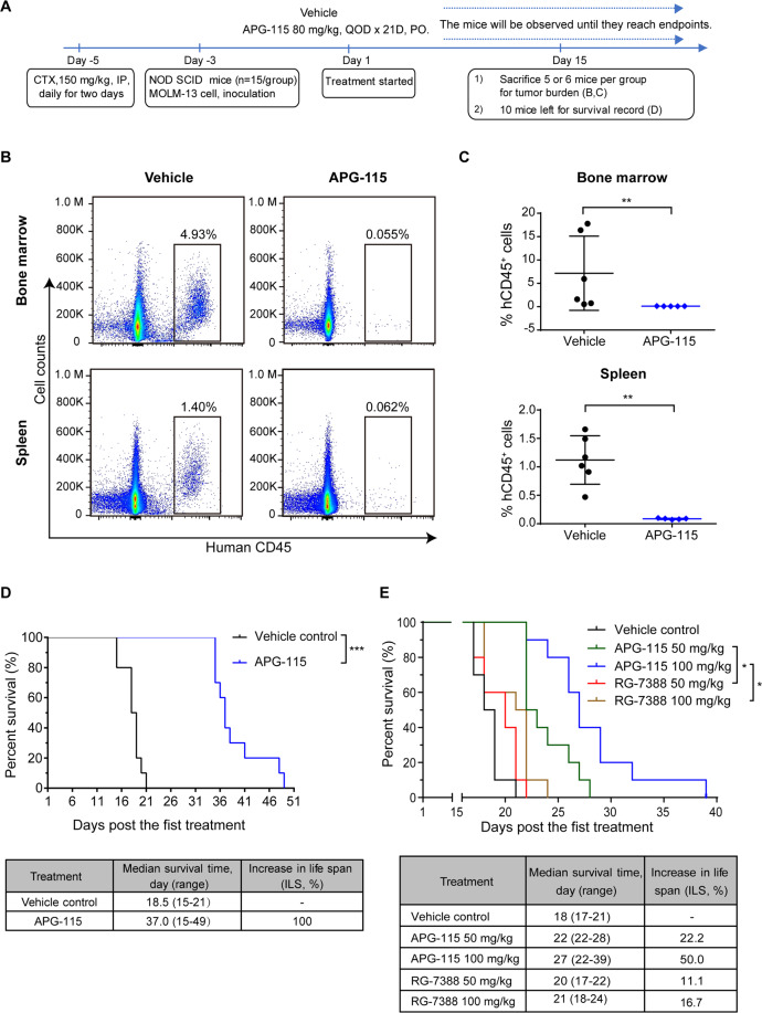
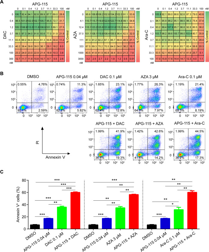
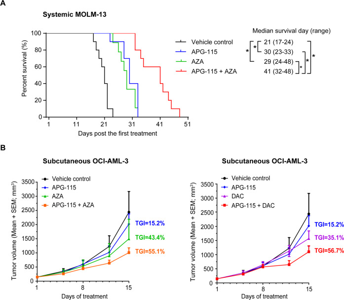
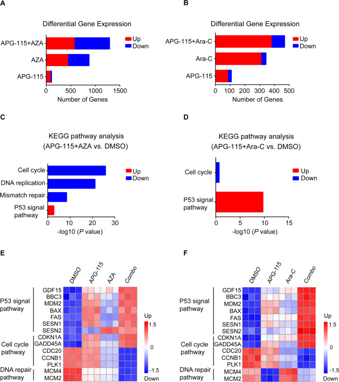
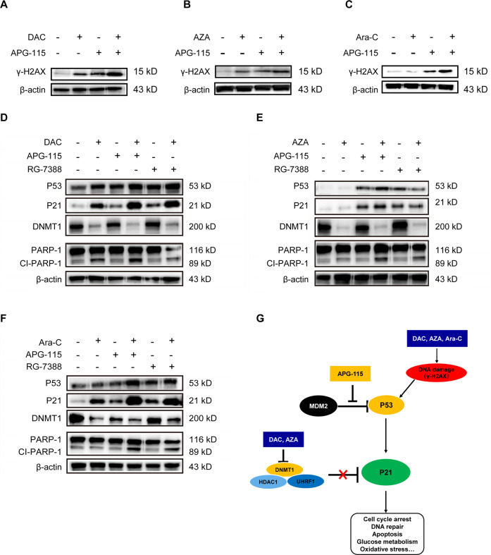
Acute myeloid leukemia (AML) is a clinically and genetically heterogeneous clonal disease associated with unmet medical needs. Paralleling the pathology of other cancers, AML tumorigenesis and propagation can be ascribed to dysregulated cellular processes, including apoptosis. This function and others are regulated by tumor suppressor P53, which plays a pivotal role in leukemogenesis. Opposing P53-mediated activities is the mouse double minute 2 homolog (MDM2), which promotes P53 degradation. Because the TP53 mutation rate is low, and MDM2 frequently overexpressed, in patients with leukemia, targeting the MDM2-P53 axis to restore P53 function has emerged as an attractive AML treatment strategy. APG-115 is a potent MDM2 inhibitor under clinical development for patients with solid tumors. In cellular cultures and animal models of AML, we demonstrate that APG-115 exerted substantial antileukemic activity, as either a single agent or when combined with standard-of-care (SOC) hypomethylating agents azacitidine (AZA) and decitabine (DAC), or the DNA-damaging agent cytarabine (Ara-C). By activating the P53/P21 pathway, APG-115 exhibited potent antiproliferative and apoptogenic activities, and induced cell cycle arrest, in TP53 wild-type AML lines. In vivo, APG-115 significantly reduced tumor burden and prolonged survival. Combinations of APG-115 with SOC treatments elicited synergistic antileukemic activity. To explain these effects, we propose that APG-115 and SOC agents augment AML cell killing by complementarily activating the P53/P21 pathway and upregulating DNA damage. These findings and the emerging mechanism of action afford a sound scientific rationale to evaluate APG-115 (with or without SOC therapies) in patients with AML.


Conflict of interest statement

The authors are full-time employees of and stockholders in Ascentage Pharma.

Figures

Fig. 1
Fig. 1. APG-115 potently inhibits cellular proliferation and induces apoptosis, with G0/G1 cell cycle arrest, in TP53wt AML cell lines.
A Mean (SD) cellular viability of MOLM-13 (TP53wt), MV-4-11 (TP53wt), OCI-AML-3 (TP53wt), HL-60 (TP53null), and SKM-1 (TP53mut) cells treated with increasing concentrations of APG-115 for 72 h (n = 3 replicates). B Total protein levels of MDM2, P53, P21, and the loading control β-actin by Western blot in MOLM-13 cells exposed to APG-115 at increasing concentrations for 4 h. C Representative flow cytometry results from two independent experiments (left panel) and mean (SD) percentage of MOLM-13 cells in apoptosis (right panel) presented as n = 4 technical replicates. *p < 0.0001; ns not significant; compared to dimethyl sulfoxide vehicle (DMSO) via two-tailed Student t tests for data from three independently repeated experiments. D Increased cell cycle arrest in the G0/G1 phase, as indicated by flow cytometry in AML cells exposed to increasing concentrations of APG-115 for 48 h. Data shown are representative images of two independent experiments.
Fig. 2
Fig. 2. Single-agent APG-115 significantly reduces the leukemic burden and prolongs survival in systemic TP53wt MOLM-13 AML xenografts.
A Study schema of the experimental paradigm to study tumor burden and animal survival in the systemic MOLM-13 AML model. For CD45 analysis, mice were treated with APG-115 for 15 days. For survival analysis, mice were treated with APG-115 for 21 days. B Representative flow cytometry results show tumor burden (i.e., engraftment of human CD45+ cells) in murine femoral bone marrow and spleen. Five or six mice treated as in A from each treatment group were euthanized and examined on Day 15. C Significantly lower proportions of human CD45+ cells in the bone marrow and spleen collected from mice exposed to APG-115 (treated as in A) compared to vehicle (**p < 0.01 by two-tailed Student tests). D Kaplan–Meier proportional-hazards plots show significantly increased percentage survival in mice receiving APG-115 per the dosing conditions shown in A (~50 days) compared to mice receiving vehicle. APG-115 prolonged median survival time by 18.5 days compared to vehicle control (37.0 vs. 18.5 days), p < 0.0001 by log-rank test; n = 9–10 mice. CTX cyclophosphamide, IP intraperitoneally, NOD SCID nonobese diabetic severe combined immunodeficient, PO orally, QOD every other day. E APG-115 confers significantly increased in vivo survival compared to RG-7388 in a systemic AML xenograft model derived from TP53 wild-type MOLM-13 cells. NOD SCID mice intravenously implanted with 1 × 107 MOLM-13 cells (n = 10/group) were treated 3 days after cell inoculation. APG-115 and RG-7388 were orally administered daily for 7 days. Kaplan–Meier curve depicts mouse survival. *p < 0.05 by log-rank test.
Fig. 3
Fig. 3. APG-115 synergizes with AZA, DAC, and Ara-C to inhibit cellular growth and induce apoptosis in TP53wt AML cells.
A Drug dose matrix of human MOLM-13 cells treated with increasing concentrations of APG-115, AZA, DAC, Ara-C or their combinations for 72 h. Cell viability was assessed using the CellTiter-Glo® assay. Representative results from three independent experiments are presented. The drug dose matrix indicates the percentage of cell viability of treated cells relative to vehicle controls. B Representative flow cytometry results from three independent experiments involving MOLM-13 cells treated with indicated concentrations of APG-115, DAC, AZA, and Ara-C (alone or in combination) for 48 h show enhanced apoptogenic effects. C Mean (SD) proportions of apoptotic cells (n = 3 replicates) show significant increases in cell lines exposed to APG-115 combined with SOC agents (vs. either alone). *p < 0.05, **p < 0.01, ***p < 0.001 by one-way ANOVA tests for data from three independently repeated experiments. DMSO, dimethylsulfoxide (vehicle); PI, propidium iodide.
Fig. 4
Fig. 4. APG-115 enhances the antileukemic activity of AZA or DAC in AML xenograft models.
Three days after cell implantation, mice were treated with APG-115 (50 mg/kg, PO, QD), AZA (2 mg/kg, IV, QD), the combination, or vehicle for 7 days. A Kaplan–Meier curves show that APG-115 combined with AZA conferred significantly prolonged survival compared to any single agent in a systemic MOLM-13 AML xenograft model of NOD SCID mice (n = 10 per group): 41 days with APG-115-AZA compared to 30 days with APG-115, 29 days with AZA, and 21 days with the vehicle (*p < 0.05 for each comparison of APG-115‒Aza with single agents or vehicle by log-rank test). B APG-115 potentiates the growth-inhibitory effects of AZA and DAC in NOD SCID mice bearing subcutaneous OCI-AML-3 xenograft tumors. Mice were treated with APG-115 (50 mg/kg, PO, QOD for 15 days), AZA (2 mg/kg, IV, QD for 7 days), DAC (1 mg/kg, IV, QD for 7 days), or combinations as indicated (n = 5 per group). Tumor growth inhibition (TGI) was calculated at the endpoint.
Fig. 5
Fig. 5. RNA sequence analysis of MOLM-13 cells treated with APG-115, AZA, Ara-C, or combinations as indicated for 24 h.
MOLM-13 cells treated with APG-115 (40 nM), AZA (3 μM), Ara-C (100 nM), alone or in combination for 24 h in triplicates. Cells were collected for RNAseq analysis after treatment. A, B Numbers of differentially expressed genes after each treatment. C, D The most significantly enriched biological processes upregulated or downregulated in response to combined treatments: both AZA and Ara-C together with APG-115 upregulated P53, while the APG-115-AZA combination more markedly downregulated AML cell cycle, DNA replication, and mismatch repair. E, F Representative differentially expressed genes in response to APG-115, AZA, or Ara-C, alone or in combination. Combinations of APG-115 with either agent upregulated genes encoding the P53 pathway and downregulated those related to the cell cycle pathway, but only the APG-115-AZA combination markedly downregulated genes implicated in DNA repair. Expression levels are presented as normalized log2 counts per million. BBC3 Bcl-2-binding component 3 gene, CCNB1 Cdc2-cyclin B1 gene, DMSO dimethylsulfoxide vehicle, CDC20 cell division cycle 20 gene, CDKN1A cyclin-dependent kinase inhibitor 1A gene, GADD45 growth arrest and DNA damage-inducible 45, GDF15 growth differentiation factor 15 gene, KEGG Kyoto Encyclopedia of Genes and Genomes, MCM2/4 minichromosome maintenance proteins 2/4 gene, PLK-1 serine/threonine-protein kinase (also termed Polo-like kinase 1) gene, SESN1/2, sestrin 1 and 2 genes.
Fig. 6
Fig. 6. Combined treatment with APG-115 and AZA, DAC, or Ara-C coordinately induces DNA damage and activates the P53/P21 pathway in AML cells.
A, B MOLM-13 cells were treated with DAC (100 nM) or AZA (0.33 µM) for 24 h and then refreshed with DAC or AZA combined with APG-115 (40 nM) for an additional 24 h. Expression levels of γ-H2AX, a biomarker for DNA double-strand breaks, were assessed by Western blot. C Expression levels of γ-H2AX in MOLM-13 cells treated with Ara-C (100 nM) alone or in combination with APG-115 (40 nM) for 48 h. DF Protein expression in MOLM-13 cells treated with APG-115 (40 nM), AZA (3 µM), Ara-C (100 nM), DAC (100 nM), or RG-7388 (40 nM), or their combinations as indicated for 48 h (D, F) or 6 h (E). β-actin was included as a loading control. Representative results from three independent experiments. G Proposed mechanism of action of APG-115 in combination with AZA, DAC, or Ara-C in AML cells. Cl-PARP-1, cleaved poly (ADP‒ribose) polymerase-1; DNMT1, DNA methyltransferase inhibitor; HDAC, histone deacetylase; γ-H2AX, H2A histone family member X; UHRF1, ubiquitin-like with PHD and RING finger domains 1.

References

    1. Lindsley RC, et al. Acute myeloid leukemia ontogeny is defined by distinct somatic mutations. Blood. 2015;125:1367–1376. - PMC - PubMed
    1. Grove CS, Vassiliou GS. Acute myeloid leukaemia: a paradigm for the clonal evolution of cancer? Dis. Model Mech. 2014;7:941–951. - PMC - PubMed
    1. Ossenkoppele G, Lowenberg B. How I treat the older patient with acute myeloid leukemia. Blood. 2015;125:767–773. - PubMed
    1. Klepin HD. Myelodysplastic syndromes and acute myeloid leukemia in the elderly. Clin. Geriatr. Med. 2016;32:155–173. - PMC - PubMed
    1. Ritchie EK, et al. Decitabine in patients with newly diagnosed and relapsed acute myeloid leukemia. Leuk. Lymphoma. 2013;54:2003–2007. - PMC - PubMed