. 2021 May 3;11(1):9438.
doi: 10.1038/s41598-021-88840-1.

Age-related iron accumulation and demyelination in the basal ganglia are closely related to verbal memory and executive functioning

Affiliations

Age-related iron accumulation and demyelination in the basal ganglia are closely related to verbal memory and executive functioning

Davina Biel et al. Sci Rep. .

Abstract

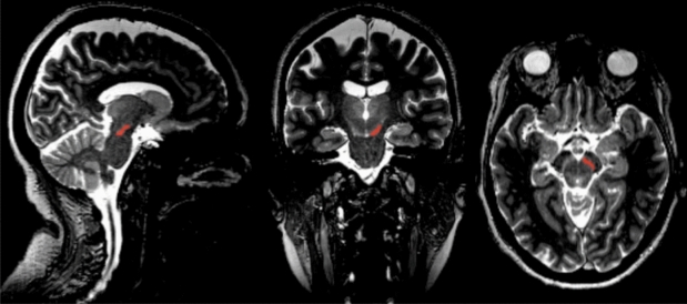
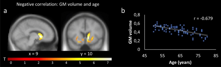
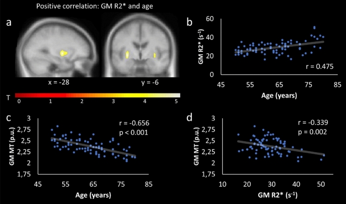
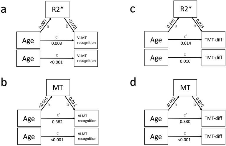
Age-related cognitive decline has been linked to alterations of the dopaminergic system and its subcortical trajectories. Recent work suggests a critical role of iron accumulation within the basal ganglia (BG) in verbal memory performance, and increased iron levels have been related to demyelination. However, the specificity of age-related iron increases with respect to cognitive functions remains unclear. Therefore, we investigated the interplay of age, cognitive performance, and structural integrity of the BG. In total, 79 healthy older participants underwent a broad cognitive assessment (fluid and crystallized intelligence, verbal and numeric memory, processing speed, executive functions) and structural MRI. As expected, performance in most cognitive tests had a negative relationship with age. Moreover, BG grey matter volume and magnetization transfer (MT, indicative of myelin) decreased, and R2* (indicative of iron) increased with age. Importantly, R2* and demyelination negatively correlated with verbal memory and executive functions. Within the SN/VTA, age correlated negatively with MT, but there was no clear evidence in favor of a relationship between behavior and R2* or MT. Our results suggest that age-related increases in iron and demyelination within the BG, which are part of a fronto-striatal network, not only impact on verbal memory but also executive functions.


Conflict of interest statement

The authors declare no competing interests.

Figures

Figure 1
Figure 1
Manual segmentation of an individual SN/VTA. Within the midbrain, the SN/VTA can be identified as bright stripe on MT-weighted images. Bilateral SN/VTAs were defined as ROIs, which were used to quantify mean MT and R2* values. For illustration purposes, only the right SN/VTA is marked in red (i.e., ROI).
Figure 2
Figure 2
Negative relationship between age and GM volume. (a) SPM showing a significant negative relationship between GM volume and age within the right caudate, pallidum, and putamen; (b) showing the corresponding correlation plot for the cluster. For display purposes, SPMs were thresholded at p < 0.001, uncorrected, and superimposed on the mean T1-weighted image.
Figure 3
Figure 3
Positive relationship between age and GM R2*. (a) SPM showing a significant positive relationship between GM R2* and age within the left and right putamen; (b) shows the corresponding correlation plot for the cluster. For post hoc analyses, GM MT values were extracted from the same cluster. They revealed a negative relationship between GM MT and age (c), and a negative relationship (not corrected for age) between GM R2* and GM MT (d). For display purposes, SPM was thresholded at p < 0.001, uncorrected, and superimposed on the mean T1-weighted image.
Figure 4
Figure 4
Relationship between cognitive performance and GM R2*. SPMs showing a significant negative relationship between GM R2* and VLMT recognition within the left and right putamen (a); a positive relationship between GM R2* and TMT within the left and right putamen (c). The corresponding correlation plots are shown in (b,d). In (d) three outliers were removed from the post hoc analysis (R2* values > 3 SD). For display purposes, SPMs were thresholded at p < 0.001, uncorrected, and superimposed on the mean T1-weighted image.
Figure 5
Figure 5
Relationship between GM MT, age, and cognitive performance. SPMs showing a significant negative relationship between GM MT and age within left and right caudate (a); a positive relationship between GM MT and VLMT recognition within the left and right caudate (c); and a negative relationship between GM MT and TMT within the left and right pallidum (e). The corresponding correlation plots for the clusters are shown in (b,d,f). For display purposes, SPMs were thresholded at p < 0.001, uncorrected, and superimposed on the mean T1-weighted image.
Figure 6
Figure 6
Relationship between MT, R2*, and age within the SN/VTA. We observed a significant negative correlation between MT and age (a); but no significant correlations between (b) R2* and age or (c) MT and R2* (p > 0.05). Benjamini-Hochberg (FDR) p-value adjustment was applied.
Figure 7
Figure 7
Illustration of the mediation model and its path estimates. Age served as predictor, VLMT recognition (a,b) and TMT-diff (c,d) as dependent variables, and R2* and MT analysis as mediator. Arrows indicate the relationship between the direct pathway (c'), and the indirect pathways (a,b). The total pathway (c) reflects the correlation between the predictor and the dependent variable. P-values are displayed on the arrows.

References

    1. Hedden T, Gabrieli JDE. Insights into the ageing mind: A view from cognitive neuroscience. Nat. Rev. Neurosci. 2004;5:87–96. doi: 10.1038/nrn1323. - DOI - PubMed
    1. Daugherty AM, Haacke EM, Raz N. Striatal iron content predicts its shrinkage and changes in verbal working memory after two years in healthy adults. J. Neurosci. 2015;35:6731–6743. doi: 10.1523/JNEUROSCI.4717-14.2015. - DOI - PMC - PubMed
    1. Steiger TK, Weiskopf N, Bunzeck N. Iron Level and Myelin Content in the Ventral Striatum Predict Memory Performance in the Aging Brain. J. Neurosci. 2016;36:3552–3558. doi: 10.1523/JNEUROSCI.3617-15.2016. - DOI - PMC - PubMed
    1. Hallgren B, Sourander P. The effect of age on the non-haemin iron in the human brain. J. Neurochem. 1958;3:41–51. doi: 10.1111/j.1471-4159.1958.tb12607.x. - DOI - PubMed
    1. Bilgic B, Pfefferbaum A, Rohlfing T, Sullivan EV, Adalsteinsson E. MRI estimates of brain iron concentration in normal aging using quantitative susceptibility mapping. Neuroimage. 2012;59:2625–2635. doi: 10.1016/j.neuroimage.2011.08.077. - DOI - PMC - PubMed

Publication types