Case Reports
. 2021 Jun;42(6):745-761.
doi: 10.1002/humu.24210. Epub 2021 May 11.

Bi-allelic KARS1 pathogenic variants affecting functions of cytosolic and mitochondrial isoforms are associated with a progressive and multisystem disease

Affiliations
Case Reports

Bi-allelic KARS1 pathogenic variants affecting functions of cytosolic and mitochondrial isoforms are associated with a progressive and multisystem disease

Gerarda Cappuccio et al. Hum Mutat. 2021 Jun.

Abstract

KARS1 encodes a lysyl-transfer RNA synthetase (LysRS) that links lysine to its cognate transfer RNA. Two different KARS1 isoforms exert functional effects in cytosol and mitochondria. Bi-allelic pathogenic variants in KARS1 have been associated to sensorineural hearing and visual loss, neuropathy, seizures, and leukodystrophy. We report the clinical, biochemical, and neuroradiological features of nine individuals with KARS1-related disorder carrying 12 different variants with nine of them being novel. The consequences of these variants on the cytosol and/or mitochondrial LysRS were functionally validated in yeast mutants. Most cases presented with severe neurological features including congenital and progressive microcephaly, seizures, developmental delay/intellectual disability, and cerebral atrophy. Oculo-motor dysfunction and immuno-hematological problems were present in six and three cases, respectively. A yeast growth defect of variable severity was detected for most variants on both cytosolic and mitochondrial isoforms. The detrimental effects of two variants on yeast growth were partially rescued by lysine supplementation. Congenital progressive microcephaly, oculo-motor dysfunction, and immuno-hematological problems are emerging phenotypes in KARS1-related disorder. The data in yeast emphasize the role of both mitochondrial and cytosolic isoforms in the pathogenesis of KARS1-related disorder and supports the therapeutic potential of lysine supplementation at least in a subset of patients.

Keywords: KARS; KARS1; LysRS; lysyl-transfer RNA synthetase; mitochondrial disease.


Conflict of interest statement

All the authors declare that there are no conflicts of interests.

Figures

Figure 1
Figure 1
Subjects with biallelic KARS1 variants and their localization on the two isoforms of KARS1 protein. Pathogenic variants in bold are novel. The p.(Ala2Val) only affects the cytosolic isoform whereas all remaining variants affect both isoforms. The cytosolic isoform refers to NM_005548.2, NP_001123561.1 (597 amino acids), and the mitochondrial isoform to NM_001130089.1, NP_005539.1 (625 amino acids)
Figure 2
Figure 2
(a) Distribution of mitochondrial disease diagnostic scores among the cases herein reported. Morphology studies were not performed in any of the cases. (b) Standard deviation score (SDS) of the occipitofrontal circumference (OFC) at birth (n = 8) and at last evaluation (n = 9). OFC at birth was not available for proband 9. OFC SDS at the latest available evaluation is statistically significantly different from birth OFC (p < .004)
Figure 3
Figure 3
Brain magnetic resonance imaging (MRI) images of case 1 (a)–(g): Axial T2‐weighted images (a), (b), (f), axial FLAIR image (b), sagittal T1‐weighted images (d) and (e), and coronal T2‐weighted image (g) at 5 months (a) and (d) and 4 years (b), (c), (e)–(g). At 5 months the temporal horns and sulci were dilated but the signal of the unmyelinated white matter appeared normal (black arrows in (a)). Note the progressive thinning of the corpus callosum and of a diffuse cerebral tissue loss causing microcephaly between 5 months and 4 years (d) and (e). In the deep temporal, frontal and peritrigonal white matter some subtle hyperintensities appeared between 5 months and 4 years, with a more preserved signal in the posterior white matter (black and white arrows respectively in (b), (c), (f)). Brain MRI images and magnetic resonance spectroscopy (MRS) of case 2 at 12 months (i) and (k) and 17 months (h), (j), (l), and (m), (n): Axial T2‐weighted images (i), (j), axial SWI phase image (h), coronal and sagittal T2‐weighted images (k), (l), and (m). The first MRI scan highlighted bilateral and confluent T2 hyperintensities in basal ganglia, thalamic nuclei, and in capsular, deep, and peripheral white matter causing tissue swelling and sulcal effacement (i) and (k). Note also the prominent involvement of external capsules and of the white matter of temporal lobes (white arrows in (i) and (k)). Incomplete operculization of sylvian fissures was already present in the first MRI and such dilation worsened later (black arrows in (i) and (j)). The severe enlargement of frontal and temporal sulci and the thinning of corpus callosum due to the tissue loss are evident (black arrows in (l) and (m). There is also an arachnoid cyst located inferiorly to the vermis (black arrow in (m)). The reduction of N‐acetyl‐aspartate peak and the presence of a small peak of lactate are shown on MRS (TE 144) (p). Point‐like hypointensities related to calcifications were bilaterally located in the frontal white matter (black arrows in (h)). MRI phase and magnitude SWI images of case 8 (o)–(p)) showed calcifications in the cortex of cerebellar hemispheres (black arrows). Brain MRI images of case 9 (q)–(u)): axial T2‐weighted and FLAIR images (q) and (r), coronal and sagittal T1‐weighted images (s), (t), and (u). Note the bilateral periventricular hyperintensities on T2 and FLAIR images, posteriorly containing little cavities (white arrows in (q), (r), and (s)). Mild enlargement of left frontal subarachnoid spaces and in the superior part of the vermis is also present (asterisks in (q), (r), and (u))
Figure 4
Figure 4
(a) Aligned regions around mutated amino acids. Human mitochondrial and cytosolic LysRS were aligned with LysRS from other organisms, including mammals, birds, reptiles, amphibians, bony fishes, cartilaginous fishes, echinoderms, arthropods, mollusks, fungi, monocotyledons, and dicotyledons. Conserved amino acids are highlighted in green, semi‐conserved amino acids are highlighted in yellow, amino acids conserved only in some organisms are highlighted in red. (b) Crystal structure of of LysRS (PDB: 4dpg) was loaded into PyMOL. The anticodon binding domain is indicated in purple while the catalytic domain in yellow. (c) The amino acid residues affected by variants found in cases are shown on the three different images of KARS1‐p38 complex. (d)–(h) Crystal structure of LysRS around amino acids which are mutated. Amino acids are represented as sticks and the protein is reported as cartoon. (d) Region around Arg108 and Arg205, in red, with Asp208 in blue. (e) Region around Phe291 and Ile346, in magenta, with Ile182, Leu286, and Phe368 in green. (f) Arg348, in red, in both LysRS subunits, with Glu295 in blue. (g) Pro499 and Pro533, in the dark green, in the catalytic domain with Lys and AMP as sticks in CPK colors. (h) Phe585, in magenta, with Tyr401, Val518, and Met519 in green
Figure 5
Figure 5
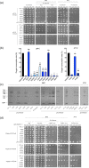
(a) Oxidative growth phenotype of haploid msk1Δ strains transformed with mtKARS1 wild‐ type or mutant alleles cloned in pFL39‐TEToff, or MSK1 cloned in pFL39. Growth assays (10‐fold dilution spots starting from 5 × 104 cells/spot) were performed in SC medium supplemented with the indicated carbon sources at 28°C and 37° C, and pictures were taken after 2–5 days of growth. The one‐letter nomenclature used in yeast has been utilized. (b) Respiratory activity of the same strains reported in (a). Oxygen consumption rate (OCR) was measured on at least four independent clones. OCR was normalized to the strain transformed with the mtKARS1 wild‐type allele. Values are means ± standard deviation. *p < .05; *** p < .001 relative to mtKARS1 wild‐type strain and # p < .05; ### p < .001 relative to empty plasmid strain using ANOVA followed by a Bonferroni's post hoc test. (c) Representative images of the mitochondrial protein synthesis of the strains reported in (a) at 28°C and 37°C. Experiments were performed on three independent clones for each mutant. (d) Viability of haploid krs1Δ strains transformed with cytKARS​1 wild‐type or mutant alleles cloned in YEplac112‐TEToff, or KRS1 cloned in pFL39. Growth assays were performed growing cells in liquid SC medium supplemented with uracil for 24 h until the early stationary phase and plating them (10‐fold dilution spots starting from 4 × 105 or 4 × 104 cells/spot) in SC medium supplemented with 2% glucose, with or without 5‐FOA, at the indicated temperatures, and pictures were taken after 2–4 days of growth

References

    1. Ardissone, A. , Tonduti, D. , Legati, A. , Lamantea, E. , Barone, R. , Dorboz, I. , Boespflug‐Tanguy, O. , Nebbia, G. , Maggioni, M. , Garavaglia, B. , Moroni, I. , Farina, L. , Pichiecchio, A. , Orcesi, S. , Chiapparini, L. , & Ghezzi, D. (2018). KARS‐related diseases: Progressive leukoencephalopathy with brainstem and spinal cord calcifications as new phenotype and a review of literature. Orphanet Journal of Rare Diseases, 13, 45. - PMC - PubMed
    1. Avula, S. , Parikh, S. , Demarest, S. , Kurz, J. , & Gropman, A. (2014). Treatment of mitochondrial disorders. Current Treatment Options in Neurology, 16, 292. - PMC - PubMed
    1. Baruffini, E. , Ferrero, I. , & Foury, F. (2010). In vivo analysis of mtDNA replication defects in yeast. Methods, 51, 426–436. - PubMed
    1. Boonsawat, P. , Joset, P. , Steindl, K. , Oneda, B. , Gogoll, L. , Azzarello‐Burri, S. , Sheth, F. , Datar, C. , Verma, I. C. , Puri, R. D. , Zollino, M. , Bachmann‐Gagescu, R. , Niedrist, D. , Papik, M. , Figueiro‐Silva, J. , Masood, R. , Zweier, M. , Kraemer, D. , Lincoln, S. , … Rauch, A. (2019). Elucidation of the phenotypic spectrum and genetic landscape in primary and secondary microcephaly. Genetics in Medicine, 21, 2043–2058. - PMC - PubMed
    1. Carmi‐Levy, I. , Motzik, A. , Ofir‐Birin, Y. , Yagil, Z. , Yang, C. M. , Kemeny, D. M. , Han, J. M. , Kim, S. , Kay, G. , Nechushtan, H. , Suzuki, R. , Rivera, J. , & Razin, E. (2011). Importin beta plays an essential role in the regulation of the LysRS‐Ap(4)A pathway in immunologically activated mast cells. Molecular and Cellular Biology, 31, 2111–2121. - PMC - PubMed

Publication types

MeSH terms