. 2021 Dec;22(12):1464-1480.
doi: 10.1111/mpp.13067. Epub 2021 May 4.

Common bacterial blight of bean: a model of seed transmission and pathological convergence

Affiliations

Common bacterial blight of bean: a model of seed transmission and pathological convergence

Nicolas W G Chen et al. Mol Plant Pathol. 2021 Dec.

Abstract

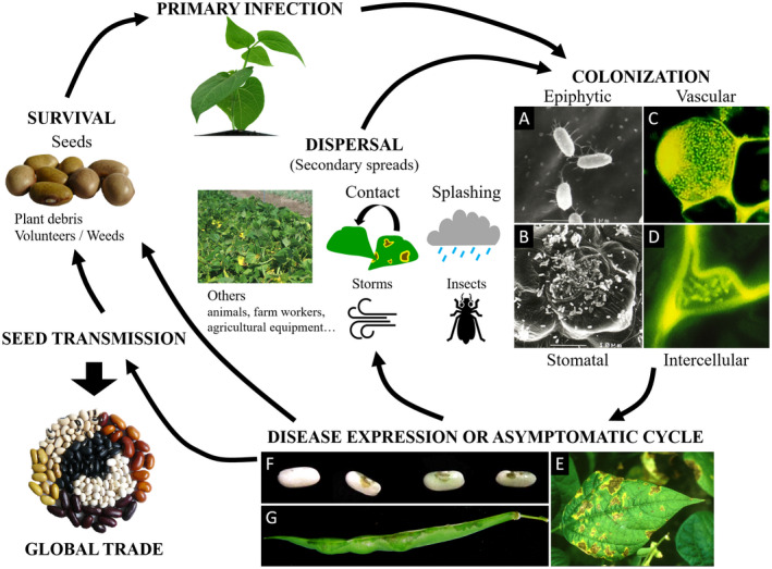
Background: Xanthomonas citri pv. fuscans (Xcf) and Xanthomonas phaseoli pv. phaseoli (Xpp) are the causal agents of common bacterial blight of bean (CBB), an important disease worldwide that remains difficult to control. These pathogens belong to distinct species within the Xanthomonas genus and have undergone a dynamic evolutionary history including the horizontal transfer of genes encoding factors probably involved in adaptation to and pathogenicity on common bean. Seed transmission is a key point of the CBB disease cycle, favouring both vertical transmission of the pathogen and worldwide distribution of the disease through global seed trade.

Taxonomy: Kingdom: Bacteria; phylum: Proteobacteria; class: Gammaproteobacteria; order: Lysobacterales (also known as Xanthomonadales); family: Lysobacteraceae (also known as Xanthomonadaceae); genus: Xanthomonas; species: X. citri pv. fuscans and X. phaseoli pv. phaseoli (Xcf-Xpp).

Host range: The main host of Xcf-Xpp is the common bean (Phaseolus vulgaris). Lima bean (Phaseolus lunatus) and members of the Vigna genus (Vigna aconitifolia, Vigna angularis, Vigna mungo, Vigna radiata, and Vigna umbellata) are also natural hosts of Xcf-Xpp. Natural occurrence of Xcf-Xpp has been reported for a handful of other legumes such as Calopogonium sp., Pueraria sp., pea (Pisum sativum), Lablab purpureus, Macroptilium lathyroides, and Strophostyles helvola. There are conflicting reports concerning the natural occurrence of CBB agents on tepary bean (Phaseolus acutifolius) and cowpea (Vigna unguiculata subsp. unguiculata).

Symptoms: CBB symptoms occur on all aerial parts of beans, that is, seedlings, leaves, stems, pods, and seeds. Symptoms initially appear as water-soaked spots evolving into necrosis on leaves, pustules on pods, and cankers on twigs. In severe infections, defoliation and wilting may occur.

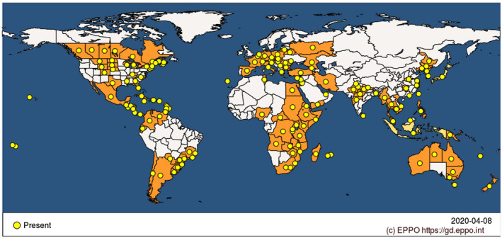
Distribution: CBB is distributed worldwide, meaning that it is frequently encountered in most places where bean is cultivated in the Americas, Asia, Africa, and Oceania, except for arid tropical areas. Xcf-Xpp are regulated nonquarantine pathogens in Europe and are listed in the A2 list by the European and Mediterranean Plant Protection Organization (EPPO).

Genome: The genome consists of a single circular chromosome plus one to four extrachromosomal plasmids of various sizes, for a total mean size of 5.27 Mb with 64.7% GC content and an average predicted number of 4,181 coding sequences.

Disease control: Management of CBB is based on integrated approaches that comprise measures aimed at avoiding Xcf-Xpp introduction through infected seeds, cultural practices to limit Xcf-Xpp survival between host crops, whenever possible the use of tolerant or resistant bean genotypes, and chemical treatments, mainly restricted to copper compounds. The use of pathogen-free seeds is essential in an effective management strategy and requires appropriate sampling, detection, and identification methods. USEFUL WEBSITES: https://gd.eppo.int/taxon/XANTPH, https://gd.eppo.int/taxon/XANTFF, and http://www.cost.eu/COST_Actions/ca/CA16107.

Keywords: Phaseolus vulgaris; Xanthomonas; common bacterial blight of bean.


Figures

FIGURE 1
FIGURE 1
Phylogeny of common bacterial blight of bean (CBB) agents. (a) Phylogenetic tree based on the core genome of Xanthomonas, constructed with the PanX pipeline (Ding et al., 2018). Briefly, 1,335 single‐copy core genes were aligned and variable positions were extracted to construct a core‐genome SNP matrix. The SNP matrix was then used to build a phylogenetic tree using FastTree2 (Price et al., 2010), which was further refined by RaxML v. 8 (Stamatakis, 2014). Arrows represent the main horizontal gene transfer events detected between Xanthomonas citri pv. fuscans (Xcf) and Xanthomonas phaseoli pv. phaseoli (Xpp) ancestors. Rooting was done using Xanthomonas vasicola strain NCPPB702, Xanthomonas campestris strain NCPPB4381, and Xanthomonas oryzae strains KACC10331, MAFF 311018, PXO99A, BAI15, BAI20, BAI21, and BLS256 (not shown) as an outgroup. (b, c) Subtrees of rep‐PCR group 9.6, from trees constructed using CVtree (Qi et al., 2004) on the same strain dataset as for the tree presented in (a). These trees are based on the frequency of appearance of overlapping oligopeptides of length K = 6. The tree in (b) was constructed based on the same 1,335 core genes as in (a), while the tree in (c) was constructed based on the pan‐genome consisting of all predicted coding sequences. Topologies in (a) and (b) are congruent with each other
FIGURE 2
FIGURE 2
Worldwide distribution of common bacterial blight of bean (CBB) agents. The distribution includes both Xanthomonas citri pv. fuscans and Xanthomonas phaseoli pv. phaseoli. Modified from https://gd.eppo.int/taxon/XANTPH/distribution (EPPO, 2021)
FIGURE 3
FIGURE 3
Common bacterial blight of bean (CBB) disease cycle and symptoms. Primary infection occurs mainly by vertical transmission through contamined seeds or from infected plant debris, volunteers, or weeds. Colonization starts with an epiphytic stage where bacteria multiply and aggregate into biofilms, followed by entry through openings such as stomata, hydathodes, or wounds. Bacteria colonize the host tissues and multiply in both the intercellular space and the vessels. Symptoms can occur on all aerial parts of the plant. Secondary spreads occur by direct contact and splashing or transport by storms and insects. Long‐distance transmission usually results from global seed trade. (a–d) Microscopy images of bacterial colonization of common bean. (a) Scanning electron microscopy (SEM) micrograph of solitary cells of epiphytic Xanthomonas citri pv. fuscans (Xcf) on bean leaves. (b) SEM micrograph of Xcf cells aggregated in biofilm in open stomata, the guard and neighbouring cells of which are largely colonized by epiphytic cells. (c) Confocal laser‐scanning microscopy (CLSM) image showing green fluorescent protein (GFP)‐tagged cells of Xcf aggregated in a biofilm adhering to the inner surface of a xylem vessel in the stem of a bean plantlet and plugging the vessel. (d) CLSM image showing GFP‐tagged cells of Xcf in the intercellular space of bean leaf parenchyma. (e–g). Images of disease expression on different plant organs. (e) Typical symptoms on leaf, showing brown necrotic lesions surrounded by a yellow halo. (f) Healthy seed (left) and three symptomatic seeds (right) showing brownish symptoms at the hilum area. (g) Pod displaying greasy symptoms spreading from the pod suture
FIGURE 4
FIGURE 4
Evidence of seed transmission through the vascular and floral pathways. Tissues or structures are indicated by letters as follows: cotyledon (co), embryo bulge (eb), external side of seed (ex), funicle (fu), hilar fissure (hf), hilum (hi), hypocotyl (hy), lens (le), micropyle (mi), osteosclerides (os), plumule (pl), palisade of macrosclereids (pm), pod suture (ps), raphe (ra), radicule (rd), tracheid bar (tb), testa (te), testa parenchyma (tp). For microscopy images, we have enhanced luminosity by 40%. (a) Schematic views of the seed. (b) Transversal section of seed and funicle imaged using confocal laser‐scanning microscopy (CLSM). (c) Sagittal plane view of a seed attached by a funicle to a pod suture. Red curved arrows represent the most probable route of vascular infection for CBB agents according to the observations made (d–k), where bacteria are mainly retrieved in the testa parenchyma. (d–k) CLSM images of a green fluorescent protein (GFP)‐tagged Xanthomonas citri pv. fuscans (Xcf) strain in bean seeds (d–g). Images were generated by merging channels 488 nm and transmitted light to observe fluorescent objects within plant tissues, or under hyperspectral detector mode (excitation at 488 and 405 nm and signal reception with all channels). In some cases, autofluorescence of noncontaminated tissues can be observed. (h–k) The same images as in (d–g) under hyperspectral mode, respectively. Plant compounds fluoresce in yellow, red, or blue (probably corresponding to cell walls, chloroplasts, and amyloplasts, respectively), confirming that the green objects observed are indeed GFP‐tagged cells. Bacteria invade the funicle parenchyma, as shown (d, h) in a transversal section of the funicle or (e, i) in a frontal section of a seed at the hilum level. (f, j) Transversal section under the hilum, showing a bacterial aggregate in a tracheid‐like cell of the tracheid bar and scattered bacteria in intercellular spaces of the testa parenchyma. (g, k) Transversal section of a mature seed showing bacterial masses of GFP‐tagged cells within the testa parenchyma. (l) Frequencies of seed transmission of Xcf wild‐type strain (wt) and mutants in the type III secretion system (hrpG: Xcf::hrpG; hrcT: Xcf::hrcT; hrcTV: Xcf::hrcV) or adhesins (pilA: Xcf∆pilA; fhaB: Xcf::fhaB; xadA2: Xcf::xadA2; yapH: Xcf::yapH). Bacterial transmission to seeds through the vascular or floral pathways was tested, respectively, by spray inoculation of leaves, keeping the reproductive organs protected, or by depositing inoculum directly in the flower buds. Thirty plants per strain and treatment were harvested individually and their seeds were analysed for the presence of Xcf (data modified from histograms presented by Darsonval et al., 2008, 2009)

References

    1. Adams, M.W. , Kelly, J.D. & Saettler, A.W. (1988) A gene for resistance to common blight (Xanthomonas campestris pv. phaseoli). Annual Report of the Bean Improvement Cooperative, 31, 73–74.
    1. Aggour, A.R. , Coyne, D.P. & Vidaver, A.K. (1989) Comparison of leaf and pod disease reactions of beans (Phaseolus vulgaris L.) inoculated by different methods with strains of Xanthomonas campestris pv. phaseoli (Smith) dye. Euphytica, 43, 143–152.
    1. Akhavan, A. , Bahar, M. , Askarian, H. , Lak, M.R. , Nazemi, A. & Zamani, Z. (2013) Bean common bacterial blight: Pathogen epiphytic life and effect of irrigation practices. Springerplus, 2, 1–9. - PMC - PubMed
    1. Alavi, S.M. , Sanjari, S. , Durand, F. , Brin, C. , Manceau, C. & Poussier, S. (2008) Assessment of the genetic diversity of Xanthomonas axonopodis pv. phaseoli and Xanthomonas fuscans subsp. fuscans as a basis to identify putative pathogenicity genes and a type III secretion system of the SPI‐1 family by multiple suppression subtractive hybridizations. Applied and Environmental Microbiology, 74, 3295–3301. - PMC - PubMed
    1. Angeles‐Ramos, R. , Vidaver, A.K. & Flynn, P. (1991) Characterization of epiphytic Xanthomonas campestris pv. phaseoli and pectolytic xanthomonads recovered from symptomless weeds in the Dominican Republic. Phytopathology, 81, 677–681.

Publication types

LinkOut - more resources