. 2021 Jul;300(1):98-107.
doi: 10.1148/radiol.2021203968. Epub 2021 May 4.

In Vivo Molecular K-Edge Imaging of Atherosclerotic Plaque Using Photon-counting CT

Affiliations

In Vivo Molecular K-Edge Imaging of Atherosclerotic Plaque Using Photon-counting CT

Salim A Si-Mohamed et al. Radiology. 2021 Jul.

Abstract

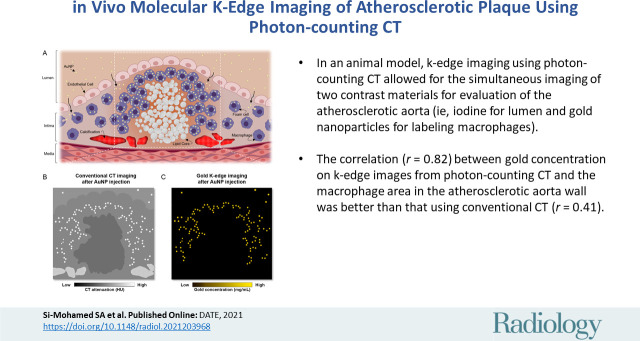
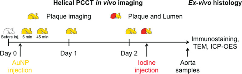
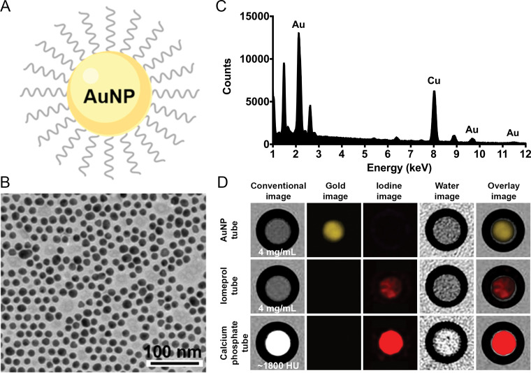
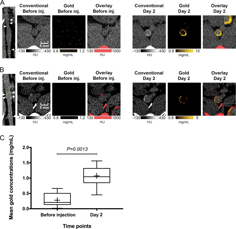
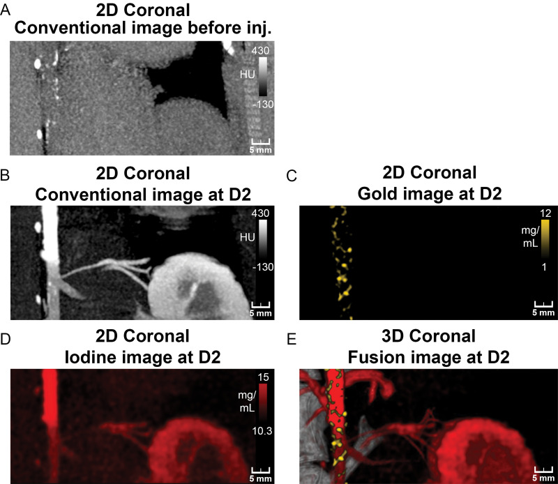
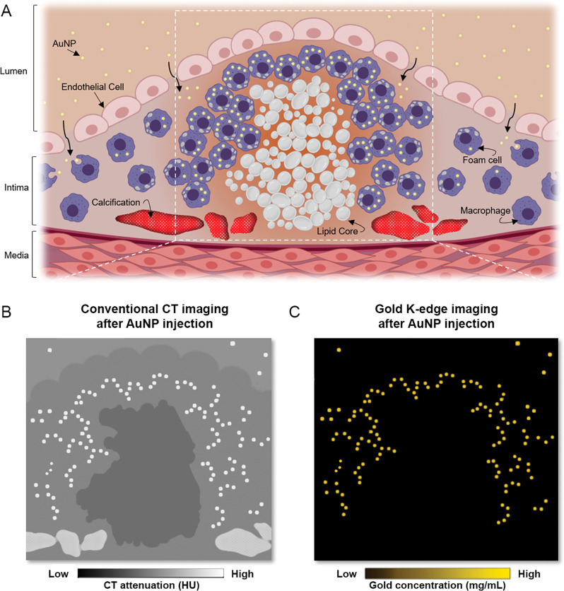
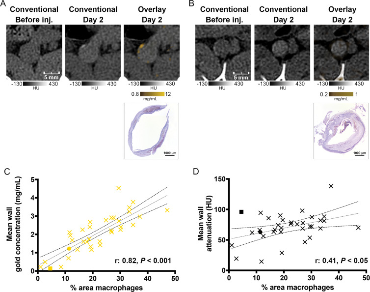
Background Macrophage burden is a major factor in the risk of atherosclerotic plaque rupture, and its evaluation remains challenging with molecular noninvasive imaging approaches. Photon-counting CT (PCCT) with k-edge imaging aims to allow for the specific detection of macrophages using gold nanoparticles. Purpose To perform k-edge imaging in combination with gold nanoparticles to detect and quantify the macrophage burden within the atherosclerotic aortas of rabbits. Materials and Methods Atherosclerotic and control New Zealand white rabbits were imaged before and at several time points up to 2 days after intravenous injection of gold nanoparticles (3.5 mL/kg, 65 mg gold per milliliter). Aortic CT angiography was performed at the end of the follow-up using an intravenous injection of an iodinated contrast material. Gold k-edge and conventional CT images were reconstructed for qualitative and quantitative assessment of the macrophage burden. PCCT imaging results were compared with findings at histologic examination, quantitative histomorphometry, transmission electron microscopy, and quantitative inductively coupled plasma optical emission spectrometry. Pearson correlations between the macrophage area measured in immunostained sections and the concentration of gold and attenuation measured in the corresponding PCCT sections were calculated. Results Seven rabbits with atherosclerosis and four control rabbits without atherosclerosis were analyzed. In atherosclerotic rabbits, calcifications were observed along the aortic wall before injection. At 2 days after injection of gold nanoparticles, only gold k-edge images allowed for the distinction of plaque enhancement within calcifications and for lumen enhancement during angiography. A good correlation was observed between the gold concentration measured within the wall and the macrophage area in 35 plaques (five per rabbit) (r = 0.82; 95% CI: 0.67, 0.91; P < .001), which was higher than that observed on conventional CT images (r = 0.41; 95% CI: 0.09, 0.65; P = .01). Transmission electron microscopy and inductively coupled plasma optical emission spectrometry analyses confirmed the gold k-edge imaging findings. Conclusion Photon-counting CT with gold nanoparticles allowed for the noninvasive evaluation of both molecular and anatomic information in vivo in rabbits with atherosclerotic plaques. Published under a CC BY 4.0 license. Online supplemental material is available for this article. See also the editorial by Leiner in this issue.


Conflict of interest statement

Disclosure of Conflicts of Interest: S.A.S.M. Activities related to the present article: disclosed no relevant relationships. Activities not related to the present article: receives payment from Boehringer for the development of educational presentations. Other relationships: disclosed no relevant relationships. M.S. disclosed no relevant relationships. J.C.H. disclosed no relevant relationships. V.T.L. disclosed no relevant relationships. L.C. disclosed no relevant relationships. P.C.N. disclosed no relevant relationships. T.G. disclosed no relevant relationships. R.D. disclosed no relevant relationships. M.C. disclosed no relevant relationships. L.B. disclosed no relevant relationships. D.P.C. Activities related to the present article: disclosed no relevant relationships. Activities not related to the present article: holds stock/stock options in PolyAurum. Other relationships: institution has patent issued. P.C.D. disclosed no relevant relationships.

Figures

None
Graphical abstract
Study design schematic. AuNP = gold nanoparticles, ICP-OES =
inductively coupled plasma optical emission spectrometry, inj. = injection,
PCCT = photon-counting CT, TEM = transmission electron
microscopy.
Figure 1:
Study design schematic. AuNP = gold nanoparticles, ICP-OES = inductively coupled plasma optical emission spectrometry, inj. = injection, PCCT = photon-counting CT, TEM = transmission electron microscopy.
In vitro characterization and imaging of gold nanoparticles (AuNP). A,
Schematic representation of a gold nanoparticle. B, Transmission electron
photomicrograph of gold nanoparticle. Note that gold nanoparticles are
represented by darker spots. C, Characteristic absorption spectrum of gold
at energy-dispersive x-ray spectroscopy. Au = gold, Cu = copper. D, In vitro
photon-counting CT images of tubes containing gold nanoparticles (4 mg/mL),
iomeprol (4 mg/mL), or calcium phosphate (1800 HU).
Figure 2:
In vitro characterization and imaging of gold nanoparticles (AuNP). A, Schematic representation of a gold nanoparticle. B, Transmission electron photomicrograph of gold nanoparticle. Note that gold nanoparticles are represented by darker spots. C, Characteristic absorption spectrum of gold at energy-dispersive x-ray spectroscopy. Au = gold, Cu = copper. D, In vitro photon-counting CT images of tubes containing gold nanoparticles (4 mg/mL), iomeprol (4 mg/mL), or calcium phosphate (1800 HU).
Photon-counting CT images of atherosclerotic rabbit aortas before and
2 days after injection (inj.) of gold nanoparticles. A, Images show
noncalcified plaque with strong circumferential enhancement and mean wall
concentration of 4.5 mg/mL of gold nanoparticles. Parietal calcifications
(arrowheads) are also shown. Green line in first image indicates aortic
section analyzed. Rectangles in Overlay Before inj. and Overlay Day 2 images
represent close-up view of parietal wall for better analyzing enhancement.
B, Images show calcified plaque with strong enhancement within and around
calcified area and mean wall concentration of 2.74 mg/mL of gold
nanoparticles. Parietal calcifications (arrowheads) are also shown. Green
line in first image indicates aortic section analyzed. Spotty areas of
enhancement were more discernable on gold k-edge image than on conventional
image. Rectangles in Overlay Before inj. and Overlay Day 2 images represent
a close-up view of parietal wall for better analyzing enhancement. C,
Box-and-whisker plot shows quantitative analysis of mean gold concentration
found in whole atherosclerotic rabbit abdominal aortic wall before injection
(mean concentration, 0.28 mg/mL ± 0.22; range, 0.01–0.66
mg/mL) and 2 days after injection (mean, 1.07 mg/mL ± 0.36; range,
0.45–1.56 mg/mL). Lower and upper margins of each box indicate 25th
and 75th percentiles. Cross represents mean, and line in box represents
median. Outliers indicate minimal and maximal values.
Figure 3:
Photon-counting CT images of atherosclerotic rabbit aortas before and 2 days after injection (inj.) of gold nanoparticles. A, Images show noncalcified plaque with strong circumferential enhancement and mean wall concentration of 4.5 mg/mL of gold nanoparticles. Parietal calcifications (arrowheads) are also shown. Green line in first image indicates aortic section analyzed. Rectangles in Overlay Before inj. and Overlay Day 2 images represent close-up view of parietal wall for better analyzing enhancement. B, Images show calcified plaque with strong enhancement within and around calcified area and mean wall concentration of 2.74 mg/mL of gold nanoparticles. Parietal calcifications (arrowheads) are also shown. Green line in first image indicates aortic section analyzed. Spotty areas of enhancement were more discernable on gold k-edge image than on conventional image. Rectangles in Overlay Before inj. and Overlay Day 2 images represent a close-up view of parietal wall for better analyzing enhancement. C, Box-and-whisker plot shows quantitative analysis of mean gold concentration found in whole atherosclerotic rabbit abdominal aortic wall before injection (mean concentration, 0.28 mg/mL ± 0.22; range, 0.01–0.66 mg/mL) and 2 days after injection (mean, 1.07 mg/mL ± 0.36; range, 0.45–1.56 mg/mL). Lower and upper margins of each box indicate 25th and 75th percentiles. Cross represents mean, and line in box represents median. Outliers indicate minimal and maximal values.
Photon-counting CT images of atherosclerotic rabbit aorta before and 2
days after injection (inj.) of gold nanoparticles. A, Coronal 5-mm-width
maximum intensity projection conventional image before injection shows
presence of focal hyperattenuation along aortic wall indicative of
calcifications. B–D, Coronal 5-mm-width maximum intensity projection
aortic CT angiograms of atherosclerotic rabbit injected with vascular
contrast material (iomerprol, 400 mg/mL) at 2 days (D2) after injection of
gold nanoparticles. Lumens of aortic and renal arteries and enhancement of
kidney and dense wall lesions are apparent on, B, maximum intensity
projection conventional and, D, iodine images. C, Coronal 5-mm-width maximum
intensity projection gold k-edge image shows extensive heterogeneous
enhancement of wall. E, Fusion of three-dimensional (3D) volume rendering of
conventional images with gold k-edge and iodine images shows spatial
distribution of atherosclerotic macrophage burden within aortic wall. 2D =
two-dimensional.
Figure 4:
Photon-counting CT images of atherosclerotic rabbit aorta before and 2 days after injection (inj.) of gold nanoparticles. A, Coronal 5-mm-width maximum intensity projection conventional image before injection shows presence of focal hyperattenuation along aortic wall indicative of calcifications. B–D, Coronal 5-mm-width maximum intensity projection aortic CT angiograms of atherosclerotic rabbit injected with vascular contrast material (iomerprol, 400 mg/mL) at 2 days (D2) after injection of gold nanoparticles. Lumens of aortic and renal arteries and enhancement of kidney and dense wall lesions are apparent on, B, maximum intensity projection conventional and, D, iodine images. C, Coronal 5-mm-width maximum intensity projection gold k-edge image shows extensive heterogeneous enhancement of wall. E, Fusion of three-dimensional (3D) volume rendering of conventional images with gold k-edge and iodine images shows spatial distribution of atherosclerotic macrophage burden within aortic wall. 2D = two-dimensional.
Molecular k-edge imaging approach using photon-counting CT in vivo for
macrophage detection and quantification in calcified atherosclerotic plaque
in combination with k-edge contrast material of gold nanoparticles (AuNP).
A, Schematic of uptake of gold nanoparticles by macrophages within
atherosclerotic plaque (gold nanoparticles are shown as yellow dots,
macrophages and foam cells are shown in purple, lipid core is shown in
white, and calcification is shown in red). Gray nodules in foam cells are
lipid droplets. White dashed lines indicate field of view of close-up in B
and C. B, Schematic of conventional CT image shows different
high-attenuation materials (gold nanoparticles and calcifications) that
hamper characterization and quantification of macrophage burden. C,
Schematic of PCCT gold k-edge image for specific, noninvasive macrophage
burden imaging, which permits quantification of gold
nanoparticles.
Figure 5:
Molecular k-edge imaging approach using photon-counting CT in vivo for macrophage detection and quantification in calcified atherosclerotic plaque in combination with k-edge contrast material of gold nanoparticles (AuNP). A, Schematic of uptake of gold nanoparticles by macrophages within atherosclerotic plaque (gold nanoparticles are shown as yellow dots, macrophages and foam cells are shown in purple, lipid core is shown in white, and calcification is shown in red). Gray nodules in foam cells are lipid droplets. White dashed lines indicate field of view of close-up in B and C. B, Schematic of conventional CT image shows different high-attenuation materials (gold nanoparticles and calcifications) that hamper characterization and quantification of macrophage burden. C, Schematic of PCCT gold k-edge image for specific, noninvasive macrophage burden imaging, which permits quantification of gold nanoparticles.
A, B, Photon-counting CT (PCCT) images of atherosclerotic macrophage
burden before injection (inj.) of gold nanoparticles and 2 days after
injection and corresponding photomicrographs obtained with immunostaining.
A, Images of noncalcified plaque show matches of spotty areas of enhancement
next to mesenteric artery branch. This section is represented by yellow dot
in C and black dot in D. B, Images of calcified plaque show low enhancement
on gold k-edge image, which is confirmed by low concentration of
immunostained macrophages. This section is represented by yellow square in C
and black square in D. C, D, Graphs show correlation between area
immunostained for macrophages and, C, concentration of gold or, D,
attenuation on conventional images. Dotted and dashed lines represent the
linear regression line and 95% CI.
Figure 6:
A, B, Photon-counting CT (PCCT) images of atherosclerotic macrophage burden before injection (inj.) of gold nanoparticles and 2 days after injection and corresponding photomicrographs obtained with immunostaining. A, Images of noncalcified plaque show matches of spotty areas of enhancement next to mesenteric artery branch. This section is represented by yellow dot in C and black dot in D. B, Images of calcified plaque show low enhancement on gold k-edge image, which is confirmed by low concentration of immunostained macrophages. This section is represented by yellow square in C and black square in D. C, D, Graphs show correlation between area immunostained for macrophages and, C, concentration of gold or, D, attenuation on conventional images. Dotted and dashed lines represent the linear regression line and 95% CI.

Comment in

References

    1. Mozaffarian D, Benjamin EJ, Go AS, et al. . Executive Summary: Heart Disease and Stroke Statistics--2016 Update: A Report From the American Heart Association. Circulation 2016;133(4):447–454. - PubMed
    1. Naghavi M, Libby P, Falk E, et al. . From vulnerable plaque to vulnerable patient: a call for new definitions and risk assessment strategies: Part I. Circulation 2003;108(14):1664–1672. - PubMed
    1. Libby P. Inflammation in atherosclerosis. Nature 2002;420(6917):868–874. - PubMed
    1. Prati F, Mallus MT, Broglia L, Albertucci M. Integrated non-invasive imaging techniques. EuroIntervention 2010;6(Suppl G):G161–G168. - PubMed
    1. Si-Mohamed S, Bar-Ness D, Sigovan M, et al. . Review of an initial experience with an experimental spectral photon-counting computed tomography system. Nucl ­Instrum Methods Phys Res A 2017;873(27):35.

Publication types

LinkOut - more resources