

Relationship Between Optical Coherence Tomography Parameter and Visual Function in Eyes With Epiretinal Membrane
- PMID: 33944891
- PMCID: PMC8107485
- DOI: 10.1167/iovs.62.6.6
Relationship Between Optical Coherence Tomography Parameter and Visual Function in Eyes With Epiretinal Membrane
Abstract
Purpose: To investigate the associations between visual function and the optical coherence tomography (OCT) parameters in eyes with idiopathic epiretinal membrane (ERM).
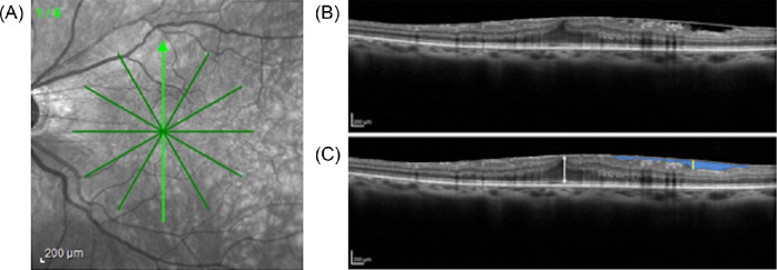
Methods: Thirty-nine consecutive eyes with ERM were enrolled. In addition to OCT parameters, such as central retinal thickness (CRT), the area of gap between the ERM and the retinal surface (SUKIMA) was newly defined and calculated from the vertical and horizontal OCT images (SUKIMAv and SUKIMAh). The average of SUKIMAv and SUKIMAh (SUKIMAave) was used for the statistical analysis. The vertical and horizontal metamorphopsia scores (MV, MH) and the average of MV and MH (Mave) were also used for the analysis.
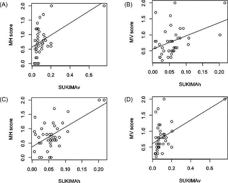
Results: The Mave was not significantly associated with logMAR visual acuity (VA) (P = 0.57, linear regression analysis). Analysis using second-order bias-corrected Akaike information criterion model selection identified the age, CRT, and SUKIMAave as being associated with logMAR VA. On the other hand, among the OCT parameters, SUKIMAave and CRT were associated with the Mave. In addition, there was a significant relationship between SUKIMAh and MV (P = 0.011) and between SUKIMAv and MH (P = 0.0014).
Conclusions: We identified SUKIMA as a novel OCT parameter that is useful to predict both VA and metamorphopsia in patients with ERM.
Conflict of interest statement
Disclosure:
Figures


Similar articles
-
Predictive factors for postoperative visual function in eyes with epiretinal membrane.Sci Rep. 2023 Dec 14;13(1):22198. doi: 10.1038/s41598-023-49689-8. Sci Rep. 2023. PMID: 38097656 Free PMC article.
-
Time course of changes in metamorphopsia, visual acuity, and OCT parameters after successful epiretinal membrane surgery.Invest Ophthalmol Vis Sci. 2012 Jun 14;53(7):3592-7. doi: 10.1167/iovs.12-9493. Invest Ophthalmol Vis Sci. 2012. PMID: 22589432
-
EN-FACE OPTICAL COHERENCE TOMOGRAPHY IN PATIENTS WITH EPIRETINAL MEMBRANE: Intuitive Method for Predicting Functional Outcomes.Retina. 2020 Oct;40(10):1972-1979. doi: 10.1097/IAE.0000000000002686. Retina. 2020. PMID: 31764613
-
THE EFFECT OF INTERNAL LIMITING MEMBRANE PEELING ON IDIOPATHIC EPIRETINAL MEMBRANE SURGERY, WITH A REVIEW OF THE LITERATURE.Retina. 2017 May;37(5):873-880. doi: 10.1097/IAE.0000000000001263. Retina. 2017. PMID: 27617536 Review.
-
Idiopathic Epiretinal Membranes - Pathophysiology, Classifications and OCT-Biomarkers.Klin Monbl Augenheilkd. 2024 May;241(5):666-674. doi: 10.1055/a-2043-4662. Epub 2023 Feb 27. Klin Monbl Augenheilkd. 2024. PMID: 36849107 Review. English, German.
Cited by
-
Superficial and Deep Capillary Plexuses: Potential Biomarkers of Focal Retinal Defects in Eyes Affected by Macular Idiopatic Epiretinal Membranes? A Pilot Study.Diagnostics (Basel). 2022 Dec 17;12(12):3205. doi: 10.3390/diagnostics12123205. Diagnostics (Basel). 2022. PMID: 36553212 Free PMC article.
-
Associations between macular retinal vasculature and severity of idiopathic epiretinal membrane.BMC Ophthalmol. 2023 May 5;23(1):200. doi: 10.1186/s12886-023-02945-x. BMC Ophthalmol. 2023. PMID: 37147577 Free PMC article.
-
Predictive factors for postoperative visual function in eyes with epiretinal membrane.Sci Rep. 2023 Dec 14;13(1):22198. doi: 10.1038/s41598-023-49689-8. Sci Rep. 2023. PMID: 38097656 Free PMC article.
-
Metamorphopsia after surgery for rhegmatogenous retinal detachment.Int J Ophthalmol. 2025 Jan 18;18(1):168-177. doi: 10.18240/ijo.2025.01.21. eCollection 2025. Int J Ophthalmol. 2025. PMID: 39829626 Free PMC article. Review.
-
Visual Performance and Predictive OCT Biomarkers in Epiretinal Membrane Assessment: Beyond Distance Visual Acuity.Invest Ophthalmol Vis Sci. 2025 Jan 2;66(1):31. doi: 10.1167/iovs.66.1.31. Invest Ophthalmol Vis Sci. 2025. PMID: 39804627 Free PMC article.
References
-
- Miyazaki M, Nakamura H, Kubo M, et al. .. Prevalence and risk factors for epiretinal membranes in a Japanese population: the Hisayama study. Graefes Arch Clin Exp Ophthalmol. 2003; 241(8): 642–646. - PubMed
-
- Kawasaki R, Wang JJ, Sato H, et al. .. Prevalence and associations of epiretinal membranes in an adult Japanese population: the Funagata study. Eye (Lond). 2009; 23(5): 1045–1051. - PubMed
-
- Fraser-Bell S, Ying-Lai M, Klein R, Varma R, Los Angeles Latino Eye Study. Prevalence and associations of epiretinal membranes in Latinos: the Los Angeles Latino Eye Study. Invest Ophthalmol Vis Sci. 2004; 45(6): 1732–1736. - PubMed
-
- Mitchell P, Smith W, Chey T, Wang JJ, Chang A. Prevalence and associations of epiretinal membranes. The Blue Mountains Eye Study, Australia. Ophthalmology . 1997; 104(6): 1033–1040. - PubMed
-
- Cheung N, Tan S-P, Lee SY, et al. .. Prevalence and risk factors for epiretinal membrane: the Singapore Epidemiology of Eye Disease study. Br J Ophthalmol. 2017; 101(6): 371–376. - PubMed
MeSH terms
LinkOut - more resources
Full Text Sources
Other Literature Sources
Medical
Research Materials
Miscellaneous
